

Short-term poor glycemic control and retinal microvascular changes in pediatric Type 1 Diabetes patients in Singapore: a pilot study
- PMID: 28615013
- PMCID: PMC5471853
- DOI: 10.1186/s12886-017-0449-8
Short-term poor glycemic control and retinal microvascular changes in pediatric Type 1 Diabetes patients in Singapore: a pilot study
Abstract
Background: Poor glycemic control in Type 1 Diabetes (T1D) patients is strongly associated with an increased risk of diabetes-related microvascular complications later in life, but it is unclear whether short period of poor glycemic control in children with T1D can cause evident microvascular morphological changes long before any pathological manifestation. Our study aimed to investigate the longitudinal association between poor glycemic control and subsequent changes in retinal microvasculature, in a pilot study of 55 pediatric T1D patients from Singapore after a one-year follow-up.
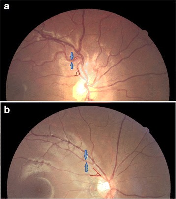
Methods: This is a hospital-based, exposure-matched and retrospective longitudinal study. A total of 55 T1D patients were included from Singapore KK Women's and Children Hospital, 28 of whom had poor glycemic control (average glycated hemoglobin [HbA1c] ≥8% during the year) while the other 27 age- and gender-matched subjects had good glycemic control (HbA1c <8%). Retinal photography was taken at diabetes annual screening and images were graded by trained graders using a semi-automated computer-based program (Singapore I Vessel Assessment [SIVA], version 4.0, Singapore Eye Research Institute, Singapore) and a spectrum of retinal vascular parameters (e.g. caliber, tortuosity, branching angle and fractal dimension) were measured quantitatively from 0.5 to 2.0 disc diameters.
Results: There was no significant difference in ethnicity, duration of T1D, blood pressure, body mass index (BMI) and low-density cholesterol lipoprotein (LDL) between the two groups. Retinal imaging was obtained at the end of 1 year of glycemic control assessment. In multiple linear regression adjusting for ethnicity, BMI, LDL and duration of T1D, patients with poor glycemic control tended to have marginally wider retinal arteriolar caliber (6.0 μm, 95% CI: -0.9, 12.8) and had significantly larger retinal arteriolar branching angle (10.1 degrees, 95% CI: 1.4, 18.9) compared with their age- and gender- matched counterparts with good glycemic control.
Conclusions: Our findings showed that abnormal retinal microvascular morphology was evident in pediatric patients with T1D after one-year's poor glycemic control. Such morphological abnormalities may lead to future development of microvascular complications among T1D pediatric patients with poor glycemic control.
Keywords: Children; Glycemic control; Retinal microvascular changes; Type 1 Diabetes (T1D).
Figures
References
-
- The effect of intensive treatment of diabetes on the development and progression of long-term complications in insulin-dependent diabetes mellitus. The Diabetes Control and Complications Trial Research Group. N Engl J Med. 1993;329(14):977–86. - PubMed
MeSH terms
Substances
LinkOut - more resources
Full Text Sources
Other Literature Sources
Medical
