. 2017 Aug 10;91(17):e00304-17.
doi: 10.1128/JVI.00304-17. Print 2017 Sep 1.

A Conserved Leucine Zipper Motif in Gammaherpesvirus ORF52 Is Critical for Distinct Microtubule Rearrangements

Affiliations

A Conserved Leucine Zipper Motif in Gammaherpesvirus ORF52 Is Critical for Distinct Microtubule Rearrangements

Matthew S Loftus et al. J Virol. .

Abstract

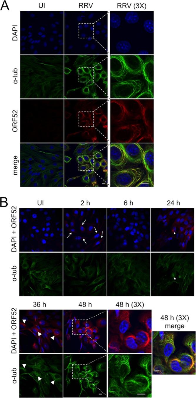
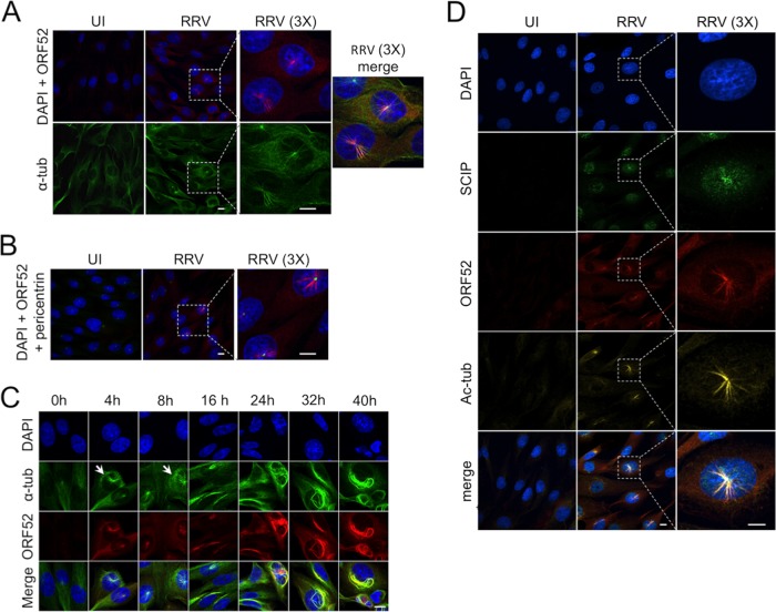
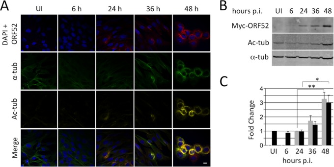
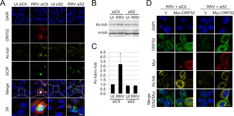
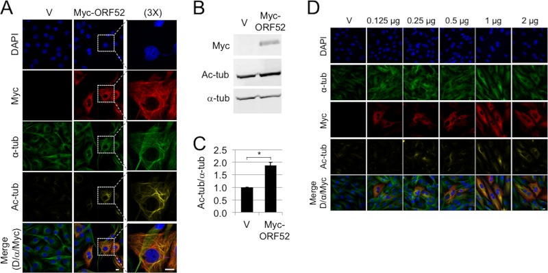
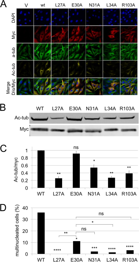
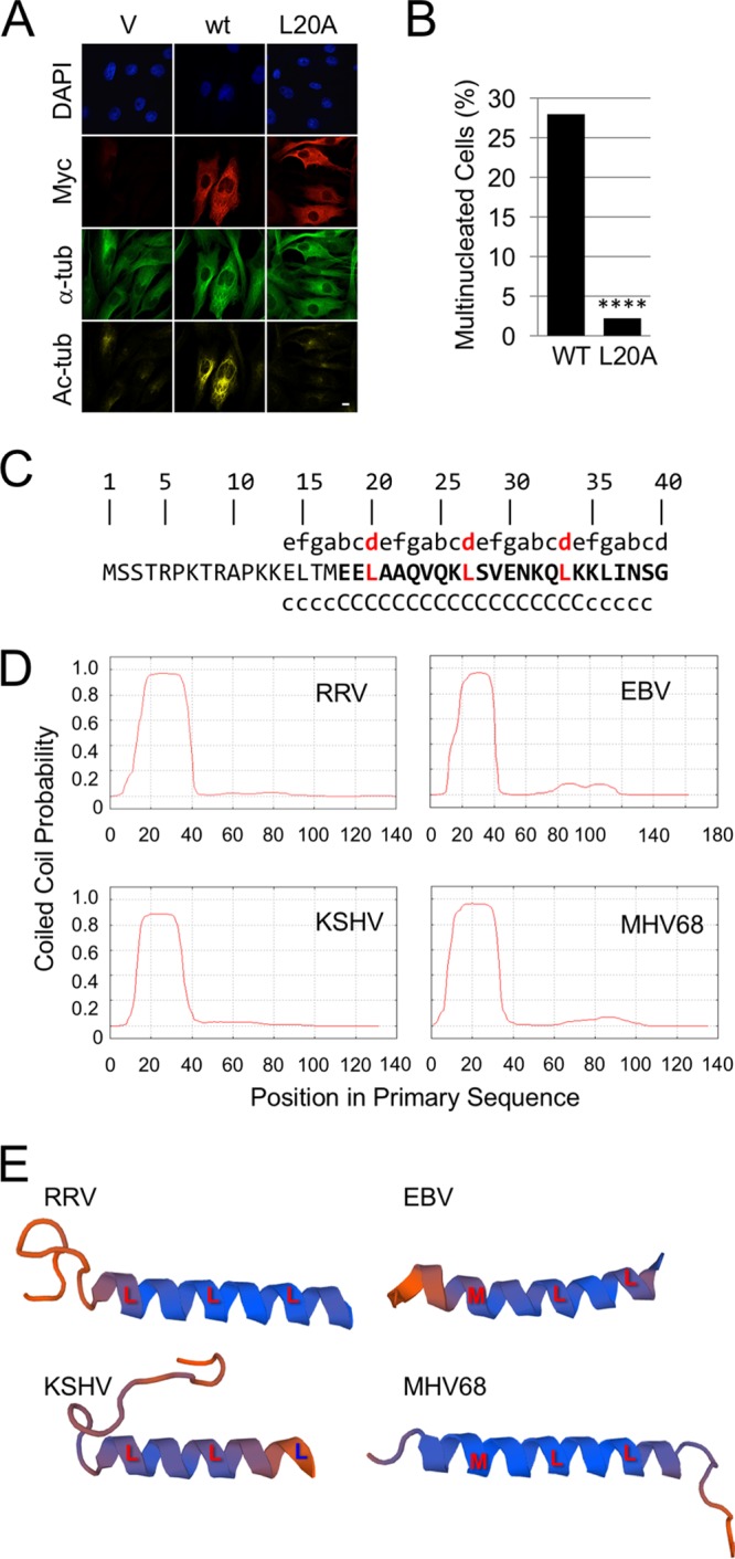
Productive viral infection often depends on the manipulation of the cytoskeleton. Herpesviruses, including rhesus monkey rhadinovirus (RRV) and its close homolog, the oncogenic human gammaherpesvirus Kaposi's sarcoma-associated herpesvirus/human herpesvirus 8 (KSHV/HHV8), exploit microtubule (MT)-based retrograde transport to deliver their genomes to the nucleus. Subsequently, during the lytic phase of the life cycle, the maturing viral particles undergo orchestrated translocation to specialized regions within the cytoplasm, leading to tegumentation, secondary envelopment, and then egress. As a result, we hypothesized that RRV might induce changes in the cytoskeleton at both early and late stages of infection. Using confocal imaging, we found that RRV infection led to the thickening and acetylation of MTs emanating from the MT-organizing center (MTOC) shortly after viral entry and more pronounced and diffuse MT reorganization during peak stages of lytic gene expression and virion production. We subsequently identified open reading frame 52 (ORF52), a multifunctional and abundant tegument protein, as being the only virally encoded component responsible for these cytoskeletal changes. Mutational and modeling analyses indicated that an evolutionarily conserved, truncated leucine zipper motif near the N terminus as well as a strictly conserved arginine residue toward the C terminus of ORF52 play critical roles in its ability to rearrange the architecture of the MT cytoskeleton. Taken together, our findings combined with data from previous studies describing diverse roles for ORF52 suggest that it likely binds to different cellular components, thereby allowing context-dependent modulation of function.IMPORTANCE A thorough understanding of the processes governing viral infection includes knowledge of how viruses manipulate their intracellular milieu, including the cytoskeleton. Altering the dynamics of actin or MT polymerization, for example, is a common strategy employed by viruses to ensure efficient entry, maturation, and egress as well as the avoidance of antiviral defenses through the sequestration of key cellular factors. We found that infection with RRV, a homolog of the human pathogen KSHV, led to perinuclear wrapping by acetylated MT bundles and identified ORF52 as the viral protein underlying these changes. Remarkably, incoming virions were able to supply sufficient ORF52 to induce MT thickening and acetylation near the MTOC, potentially aiding in the delivery viral genomes to the nucleus. Although the function of MT alterations during late stages of infection requires further study, ORF52 shares functional and structural similarities with alphaherpesvirus VP22, underscoring the evolutionary importance of MT cytoskeletal manipulations for this virus family.

Keywords: HHV8; Kaposi's sarcoma-associated herpesvirus; RRV; coiled coil; leucine zipper; microtubule-associated protein; rhesus monkey rhadinovirus; tegument.


Figures

FIG 1
FIG 1
RRV infection at a low MOI leads to MT bundling, which colocalizes with ORF52. (A) hTERT-RhF cells were mock infected (uninfected [UI]) or infected with RRV (MOI = 5) for 48 h, followed by fixation and staining for DNA (DAPI) (blue), α-tubulin (α-tub) (green), and ORF52 (red), and then imaged by confocal fluorescence microscopy. The bottom images represent the merging of the images from the three stains. The third column of images shows ×3 magnifications of the regions indicated by the dashed boxes in the adjacent images. (B) hTERT-RhF cells were infected with RRV (MOI = 5), fixed at the indicated times from 2 to 48 h p.i., and stained as described above for panel A. The top rows of images represent a merge of DAPI and ORF52 staining, and the bottom rows represent α-tubulin staining. Cells within the boxed region from the 48-h time point are shown at a ×3 magnification in the adjacent column, and the final image depicts all 3 signals merged. Arrows in the images at 2 h p.i. indicate examples of cells with low levels of ORF52 likely released from the initial RRV inoculum. Thin arrowheads in the images for 24 h p.i. indicate what appears to be an MT aster, and triangular arrowheads indicate moderate MT bundling at 36 h p.i.
FIG 2
FIG 2
RRV infection at a high MOI leads to the transient appearance of ORF52-decorated MT asters at the MTOC and centripetal capsid accumulation. (A) Mock- or RRV-infected (MOI = 50) hTERT-RhF cells were incubated for 4 h and then stained and imaged as described in the legend of Fig. 1. The third column shows magnified (×3) views of the boxed regions in the adjacent images, and the third row represents the merging of all three stains. (B) Parallel aliquots of infected hTERT-RhF cells were stained for pericentrin (green) in addition to DAPI and ORF52. (C) Additional hTERT-RhF cells were infected at a high MOI and incubated for the times indicated. Cells were then fixed, stained, and imaged as described above for panel A. The bottom row is a merge of DAPI, ORF52, and α-tubulin. Short arrows indicate examples of cells with MT asters. (D) Mock-infected (UI) or RRV-infected (MOI = 50) hTERT-RhF cells were incubated for 4 h and then stained with DAPI, SCIP (ORF65), or ORF52. The third column shows magnified (×3) views of the boxed regions in the adjacent images, and the bottom row represents the merging of all three stains. White bars represent 10 μm.
FIG 3
FIG 3
Acetylation of RRV-induced MT bundles correlates and colocalizes with ORF52 expression. (A) hTERT-RhF cells were mock infected (UI) or infected with RRV (MOI = 5) and fixed at indicated times p.i. Cells were stained for DNA (DAPI) (blue), ORF52 (red), α-tubulin (green), or acetylated tubulin (Ac-tub) (dark yellow), with the bottom row displaying merged images. (B) RRV-infected hTERT-RhF cells were collected at increasing times p.i. and analyzed for levels of ORF52 expression, tubulin acetylation, and total α-tubulin (loading control) by immunoblotting with the indicated antibodies. (C) Graphic representation of ORF52 expression (gray bars) and tubulin acetylation (black bars) from 3 biological replicates of the experiment shown in panel B. Values represent means ± standard errors of the means, normalized to the α-tubulin level for each lane. The ORF52 expression level is set to 1.0 at 24 h, and that for acetylated tubulin is set to 1.0 for uninfected cells (*, P < 0.05; **, P < 0.01 [by 2-tailed Student's t test]). All micrographs in panel A are at the same magnification, and the white bar represents 10 μm.
FIG 4
FIG 4
ORF52 is necessary for MT bundling during infection. (A) hTERT-RhF cells were transfected with siCtl or si52 and incubated for 24 h. Cells were then either mock infected (UI) or infected with RRV (MOI = 5); incubated for an additional 48 h; and then stained for DNA (blue), ORF65 (SCIP) (green), ORF52 (red), and acetylated tubulin (dark yellow). The penultimate row is a merge of all four colors in the column. The last row is a magnified (×3) view of the cells within the boxed area of the adjacent merged image. (B) Immunoblot analysis of the expression of acetylated tubulin and α-tubulin of hTERT-RhF cells treated the same way as described above for panel A. (Of note, the second lane, siCtl RRV, was underloaded.) (C) Graphic representation of the ratios of acetylated to alpha tubulin under the indicated conditions. The bars are the means ± standard errors of the means of data from three experiments, and the asterisks indicate ratios that differed significantly (P < 0.05, by Student's t test) in RRV-infected cells treated with control siRNA. (D) RRV-infected cells pretreated with siCtl or si52 were transfected with either the vector (V) or a plasmid encoding si52-resistant ORF52, as indicated. Forty hours later, the cells were fixed and stained as indicated. White bars represent 10 μm in panel A. siCtl, control siRNA; si52, siRNA against ORF52.
FIG 5
FIG 5
ORF52 is sufficient by itself to induce MT bundling. (A) Immortalized RhF cells were Amaxa transfected with Myc-tagged ORF52 or the empty Myc-tagged vector (V) and incubated for 48 h. Cells were fixed and stained with DAPI (blue), α-tubulin (green), acetylated tubulin (dark yellow), and Myc (red) and imaged by confocal microscopy. The last column of images shows cells from the boxed areas at a ×3 magnification. The bottom row is a merge of DAPI, ORF52, and alpha tubulin. (B) RhF cells were transfected with the vector alone or wt RRV ORF52 as described above for panel A and then analyzed 48 h later by immunoblotting with antibodies against acetylated tubulin and α-tubulin and Myc-tagged RRV ORF52 (Myc-ORF52). (C) Graphic representation of data from 3 replicative experiments depicted in panel B, quantifying the ratio of acetylated to total α-tubulin. Values were normalized to the value with the vector alone (pK-Myc). Asterisks indicate statistical significance (P < 0.05) determined by 2-tailed Student's t test. Error bars represent standard errors of the means. (D) Smaller amounts of transfected DNA lead to less obvious MT acetylation and bundling, but the failure of cytokinesis persists. hTERT-RhF cells were transfected with the empty Myc-tagged vector or increasing amounts of the Myc-ORF52-encoding plasmid as indicated. After 48 h, cells were fixed, stained, and imaged. The bottom row is a merge of DAPI, ORF52, and α-tubulin. The white bars represent 10 μm.
FIG 6
FIG 6
Four of the five strictly conserved residues of RRV ORF52 are important for MT bundling and acetylation. (A) hTERT-RhF cells were transfected with the pK-Myc vector, the RRV Myc-ORF52 wt, or a RRV Myc-ORF52 alanine mutant. Cells were stained with DAPI or the indicated monoclonal antibodies. The white bar indicates 10 μm. (B) hTERT-RhF cells were transfected in the same way as described above for panel A. Cells were incubated for 48 h, and cell lysates were immunoblotted with anti-acetylated tubulin and anti-Myc (Myc) antibodies. (C) Graphic representation of data from four replicative experiments depicted in panel B, quantifying the ratio of acetylated tubulin to total Myc expression. (D) Fifty to one hundred cells in randomly selected microscopy fields under each of the transfection conditions indicated in panel A were scored for single or multiple nuclei. Asterisks directly over bars in panel C indicate statistical significance determined by Student's t test, comparing the wt to the indicated mutant, and error bars represent standard errors of the means. For panel D, asterisks above bars indicate statistical significance determined by a chi-squared test, comparing the wt to the indicated mutant, and those above brackets are for comparisons of the E30A mutant with the other indicated mutants. *, **, ***, and **** represent P values of <0.05, 0.01, 0.001, and 0.0001, respectively. ns, not statistically significant.
FIG 7
FIG 7
The N-terminal region of RRV ORF52 contains a single, evolutionarily conserved heptad repeat suggestive of a truncated leucine zipper that was essential for MT bundling and acetylation. (A) IF analysis of hTERT-RhF cells 48 h after transfection with the pK-Myc vector (V), the RRV ORF52 wt (wt), or the RRV ORF52 L20A mutant. (B) Multinucleated cells were quantified as described in the legend of Fig. 5, with asterisks indicating a P value of <0.0001 as determined by a chi-squared test. (C) The first 40 amino acids of RRV ORF52. The top line represents heptad repeat positions a through g, with the critical d position shown in red. The second line is the amino acid sequence, with bold font indicating the predicted α-helix (37, 38, 42). The third line delineates the coiled-coil prediction, with c and C representing coiled-coil probabilities of >50% and >90%, respectively, using the Marcoil algorithm (37). (D) Profiles of coiled-coil predictions for RRV, KSHV, EBV, and MHV-68 ORF52 orthologs over the length of each protein, showing positional conservation. (E) Swiss-Model-predicted three-dimensional structures of the first 40 amino acids of the RRV, EBV, KSHV, and MHV-68 ORF52 orthologs, using the MHV-68 ORF52 crystal structure as the template and showing the three, heptad d-position amino acids (L or M) aligning along the same side of the α-helix.

Similar articles

Cited by

References

    1. Brice A, Whelan DR, Ito N, Shimizu K, Wiltzer-Bach L, Lo CY, Blondel D, Jans DA, Bell TDM, Moseley GW. 2016. Quantitative analysis of the microtubule interaction of rabies virus P3 protein: roles in immune evasion and pathogenesis. Sci Rep 6:33493. doi: 10.1038/srep33493. - DOI - PMC - PubMed
    1. Zhang W, Gao S-J. 2012. Exploitation of cellular cytoskeletons and signaling pathways for cell entry by Kaposi's sarcoma-associated herpesvirus and the closely related rhesus rhadinovirus. Pathogens 1:102–127. doi: 10.3390/pathogens1020102. - DOI - PMC - PubMed
    1. Diefenbach RJ, Miranda-Saksena M, Douglas MW, Cunningham AL. 2008. Transport and egress of herpes simplex virus in neurons. Rev Med Virol 18:35–51. doi: 10.1002/rmv.560. - DOI - PubMed
    1. Ogawa-Goto K, Tanaka K, Gibson W, Moriishi E, Miura Y, Kurata T, Irie S, Sata T. 2003. Microtubule network facilitates nuclear targeting of human cytomegalovirus capsid. J Virol 77:8541–8547. doi: 10.1128/JVI.77.15.8541-8547.2003. - DOI - PMC - PubMed
    1. Sodeik B. 2000. Mechanisms of viral transport in the cytoplasm. Trends Microbiol 8:465–472. doi: 10.1016/S0966-842X(00)01824-2. - DOI - PubMed

Publication types