

Modeling Physiological Events in 2D vs. 3D Cell Culture
- PMID: 28615311
- PMCID: PMC5545611
- DOI: 10.1152/physiol.00036.2016
Modeling Physiological Events in 2D vs. 3D Cell Culture
Abstract
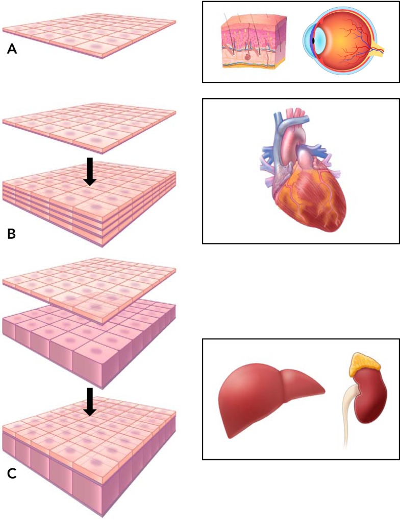
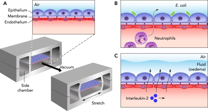
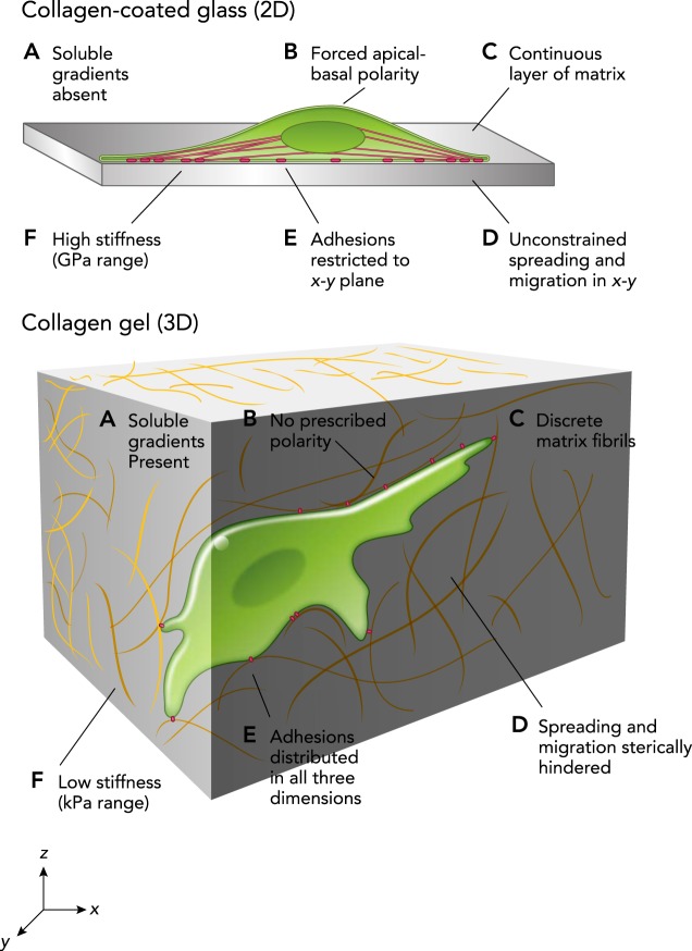
Cell culture has become an indispensable tool to help uncover fundamental biophysical and biomolecular mechanisms by which cells assemble into tissues and organs, how these tissues function, and how that function becomes disrupted in disease. Cell culture is now widely used in biomedical research, tissue engineering, regenerative medicine, and industrial practices. Although flat, two-dimensional (2D) cell culture has predominated, recent research has shifted toward culture using three-dimensional (3D) structures, and more realistic biochemical and biomechanical microenvironments. Nevertheless, in 3D cell culture, many challenges remain, including the tissue-tissue interface, the mechanical microenvironment, and the spatiotemporal distributions of oxygen, nutrients, and metabolic wastes. Here, we review 2D and 3D cell culture methods, discuss advantages and limitations of these techniques in modeling physiologically and pathologically relevant processes, and suggest directions for future research.
Copyright © 2017 the American Physiological Society.
Conflict of interest statement
No conflicts of interest, financial or otherwise, are declared by the author(s).
Figures
Comment in
-
Findings of Research Misconduct.Fed Regist. 2022 Mar 11;87(48):14020-14021. Fed Regist. 2022. PMID: 35295230 Free PMC article. No abstract available.
References
-
- Amanullah A, Otero JM, Mikola M, Hsu A, Zhang J, Aunins J, Schreyer HB, Hope JA, Russo AP. Novel micro-bioreactor high throughput technology for cell culture process development: Reproducibility and scalability assessment of fed-batch CHO cultures. Biotechnol Bioeng 106: 57–67, 2010. doi: 10.1002/bit.22664. - DOI - PubMed
-
- Beauchamp P, Moritz W, Kelm JM, Ullrich ND, Agarkova I, Anson BD, Suter TM, Zuppinger C. Development and characterization of a scaffold-free 3D spheroid model of induced pluripotent stem cell-derived human cardiomyocytes. Tissue Eng Part C Methods 21: 852–861, 2015. doi: 10.1089/ten.tec.2014.0376. - DOI - PubMed
Publication types
MeSH terms
Grants and funding
LinkOut - more resources
Full Text Sources
Other Literature Sources