Review
. 2017 Jul;32(4):278-289.
doi: 10.1152/physiol.00005.2017.

Stem Cells in Repair of Gastrointestinal Epithelia

Affiliations
Review

Stem Cells in Repair of Gastrointestinal Epithelia

Amanda Andersson-Rolf et al. Physiology (Bethesda). 2017 Jul.

Abstract

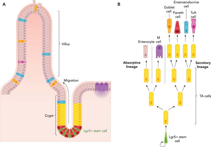
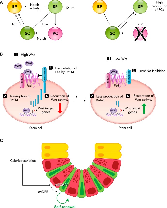
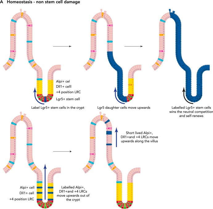
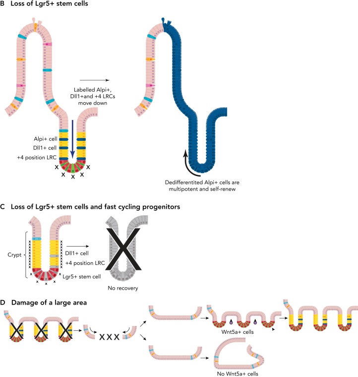
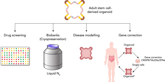
Among the endodermal tissues of adult mammals, the gastrointestinal (GI) epithelium exhibits the highest turnover rate. As the ingested food moves along the GI tract, gastric acid, digestive enzymes, and gut resident microbes aid digestion as well as nutrient and mineral absorption. Due to the harsh luminal environment, replenishment of new epithelial cells is essential to maintain organ structure and function during routine turnover and injury repair. Tissue-specific adult stem cells in the GI tract serve as a continuous source for this immense regenerative activity. Tissue homeostasis is achieved by a delicate balance between gain and loss of cells. In homeostasis, temporal tissue damage is rapidly restored by well-balanced tissue regeneration, whereas prolonged imbalance may result in diverse pathologies of homeostasis and injury repair. Starting with a summary of the current knowledge of GI tract homeostasis, we continue with providing models of acute injury and chronic diseases. Finally, we will discuss how primary organoid cultures allow new insights into the mechanisms of homeostasis, injury repair, and disease, and how this novel 3D culture system has the potential to translate into the clinic.


Conflict of interest statement

No conflicts of interest, financial or otherwise, are declared by the author(s).

Figures

FIGURE 1.
FIGURE 1.
Architecture of the small intestine A: intestinal Lgr5+ stem cells (green) are located at the bottom of the intestinal crypt in between Paneth (niche) cells (magenta). Following stem cell division, the progenitors migrate upward in a conveyor belt-like manner to undergo a few rounds of rapid division before differentiating into functional cell types. On reaching the tip of the villi, the terminally differentiated cells are shed into the lumen. B: schematic drawing illustrating how division of Lgr5+ stem cells give rise to progenitor transit amplifying (TA) cells, which then differentiate into either the secretory or absorptive lineage.
FIGURE 2.
FIGURE 2.
Signaling in intestinal stem cells A: Notch signaling in homeostasis (left) and in the absence of Paneth cells (right). Notch signaling plays a crucial role in stem cell (SC) maintenance and is supplied by the Paneth (niche) cells (PC). In the presence of Paneth cells (left), high levels of Notch signaling promote the daughter cell to adopt an absorptive progenitor fate (EP; enterocyte progenitor), whereas low levels of Notch signaling facilitate the formation of secretory progenitors (SP), some of which will differentiate into Paneth or enteroendocrine cells. In the absence of Paneth cells (right), the lack of Notch signaling promotes intestinal SC differentiation toward the secretory lineage, thus facilitating the generation of new Paneth cells. B: high (left) and low (right) levels of Wnt signaling. Left: high levels of Wnt signaling (1) leads to transcription of Rnf43 (a Wnt target gene; 2), which degrades the Wnt receptor Frizzled (3) and subsequently reduces the level of Wnt activity (4). Right: during low levels of Wnt signaling (1), the production of Rnf43 is reduced (2), and consequently there is less to no degradation of Frizzled (3), and the Wnt activity is enhanced. C: calorie restriction promotes intestinal stem cell renewal. Calorie restriction inhibits mTOR complex 1 (mTORC1) signaling in Paneth cells, resulting in the production of cyclic ADP ribose (cADPR). cADPR acts as a paracrine factor and promotes stem cell division.
FIGURE 3.
FIGURE 3.
Plasticity of the intestinal epithelium upon damage A: during homeostasis, the Lgr5+ stem cells (top) self-renew and give rise to all different cell types of the intestinal epithelium. Thus, at a later time point following labeling of an Lgr5+ stem cell, the whole crypt and villus will consist of its progeny (as long as the labeled Lgr5+ stem cell wins the “neutral competition” against the other unlabelled stem cell clones). Label-retaining cells (LRCs), Dll1+ progenitors, and Alpi+ progenitors migrate upward along the crypt villus axis under homeostatic conditions (bottom). They differentiate and are eventually lost; hence no tracing can be observed from these labeled cells. B: upon loss of Lgr5+ stem cells, LRCs, Dll1+ progenitors, and Alpi+ progenitors can dedifferentiate into a stem cell-like state and replace the lost Lgr5+ stem cells. C: upon loss of Lgr5+ stem and progenitor cells as well as rapidly dividing Lgr5- progenitors, the intestinal epithelium cannot recover. D: following loss of multiple crypts in the colonic epithelium, cells originating from the adjacent tissue migrate to quickly seal the wound. Then, the presence of Wnt5a-positive cells (purple circles) in the underlying mesenchyme stimulate crypt regeneration. In the absence of Wnt5a+ cells, no new crypts are formed.
FIGURE 3.
FIGURE 3.
Plasticity of the intestinal epithelium upon damage A: during homeostasis, the Lgr5+ stem cells (top) self-renew and give rise to all different cell types of the intestinal epithelium. Thus, at a later time point following labeling of an Lgr5+ stem cell, the whole crypt and villus will consist of its progeny (as long as the labeled Lgr5+ stem cell wins the “neutral competition” against the other unlabelled stem cell clones). Label-retaining cells (LRCs), Dll1+ progenitors, and Alpi+ progenitors migrate upward along the crypt villus axis under homeostatic conditions (bottom). They differentiate and are eventually lost; hence no tracing can be observed from these labeled cells. B: upon loss of Lgr5+ stem cells, LRCs, Dll1+ progenitors, and Alpi+ progenitors can dedifferentiate into a stem cell-like state and replace the lost Lgr5+ stem cells. C: upon loss of Lgr5+ stem and progenitor cells as well as rapidly dividing Lgr5- progenitors, the intestinal epithelium cannot recover. D: following loss of multiple crypts in the colonic epithelium, cells originating from the adjacent tissue migrate to quickly seal the wound. Then, the presence of Wnt5a-positive cells (purple circles) in the underlying mesenchyme stimulate crypt regeneration. In the absence of Wnt5a+ cells, no new crypts are formed.
FIGURE 4.
FIGURE 4.
Uses and applications of adult stem cell-derived organoids The potential uses and applications of adult stem cell-derived organoids are many. For example, they can be used for drug screening of new and already existing drugs, and be cryopreserved to form a biobank that can be used for the purpose of basic science and translational medicine. Moreover, they can be used for disease modeling, either by gene editing or by derivation from human patients. In the future, they might have the potential to be used in personalized gene therapy, e.g., correction of the genetic defect in patient-derived organoids, and then transplantation back into the patient. *Genetic defect has been corrected.

References

    1. Adolph TE, Tomczak MF, Niederreiter L, Ko H-J, Böck J, Martinez-Naves E, Glickman JN, Tschurtschenthaler M, Hartwig J, Hosomi S, Flak MB, Cusick JL, Kohno K, Iwawaki T, Billmann-Born S, Raine T, Bharti R, Lucius R, Kweon M-N, Marciniak SJ, Choi A, Hagen SJ, Schreiber S, Rosenstiel P, Kaser A, Blumberg RS. Paneth cells as a site of origin for intestinal inflammation. Nature 503: 272–276, 2013. doi: 10.1038/nature12599. - DOI - PMC - PubMed
    1. Ahn S-H, Shah YM, Inoue J, Morimura K, Kim I, Yim S, Lambert G, Kurotani R, Nagashima K, Gonzalez FJ, Inoue Y. Hepatocyte nuclear factor 4alpha in the intestinal epithelial cells protects against inflammatory bowel disease. Inflamm Bowel Dis 14: 908–920, 2008. doi: 10.1002/ibd.20413. - DOI - PMC - PubMed
    1. Andreu P, Peignon G, Slomianny C, Taketo MM, Colnot S, Robine S, Lamarque D, Laurent-Puig P, Perret C, Romagnolo B. A genetic study of the role of the Wnt/beta-catenin signalling in Paneth cell differentiation. Dev Biol 324: 288–296, 2008. doi: 10.1016/j.ydbio.2008.09.027. - DOI - PubMed
    1. Bach SP, Renehan AG, Potten CS. Stem cells: the intestinal stem cell as a paradigm. Carcinogenesis 21: 469–476, 2000. doi: 10.1093/carcin/21.3.469. - DOI - PubMed
    1. Barker N, van Es JH, Kuipers J, Kujala P, van den Born M, Cozijnsen M, Haegebarth A, Korving J, Begthel H, Peters PJ, Clevers H. Identification of stem cells in small intestine and colon by marker gene Lgr5. Nature 449: 1003–1007, 2007. doi: 10.1038/nature06196. - DOI - PubMed

Publication types