. 2017 Jun 14;9(394):eaal4508.
doi: 10.1126/scitranslmed.aal4508.

Donor pulmonary intravascular nonclassical monocytes recruit recipient neutrophils and mediate primary lung allograft dysfunction

Affiliations

Donor pulmonary intravascular nonclassical monocytes recruit recipient neutrophils and mediate primary lung allograft dysfunction

Zhikun Zheng et al. Sci Transl Med. .

Abstract

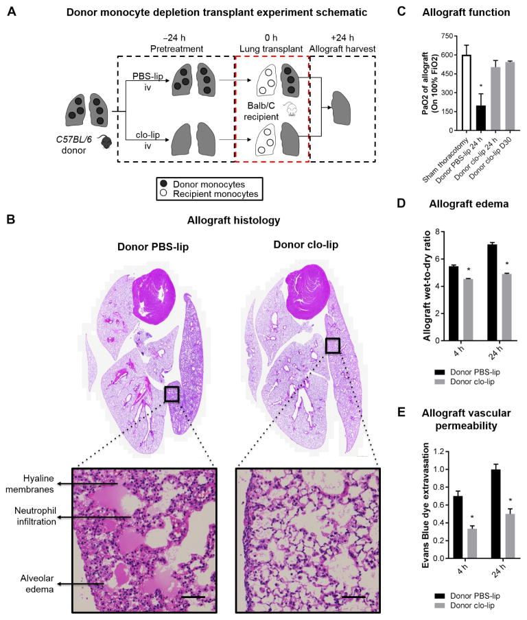
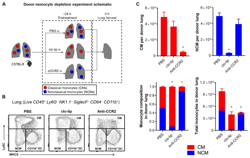
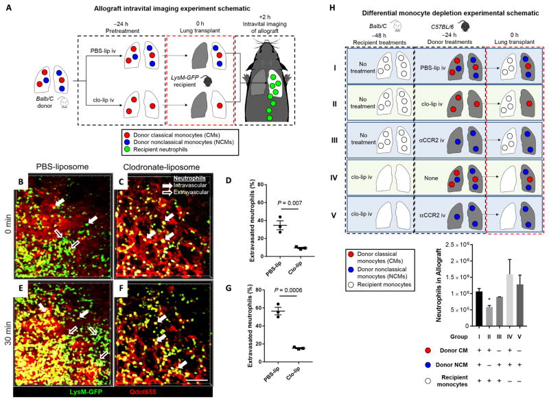
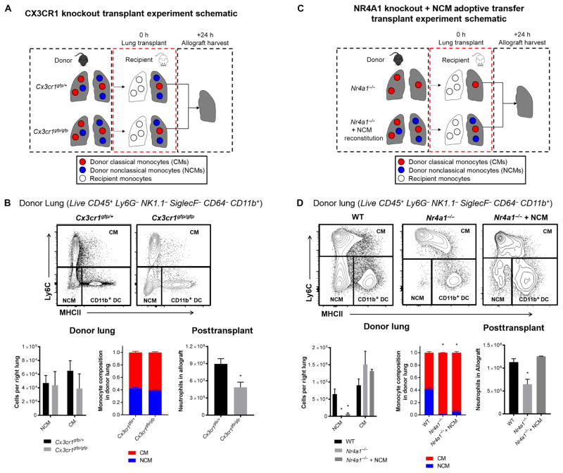
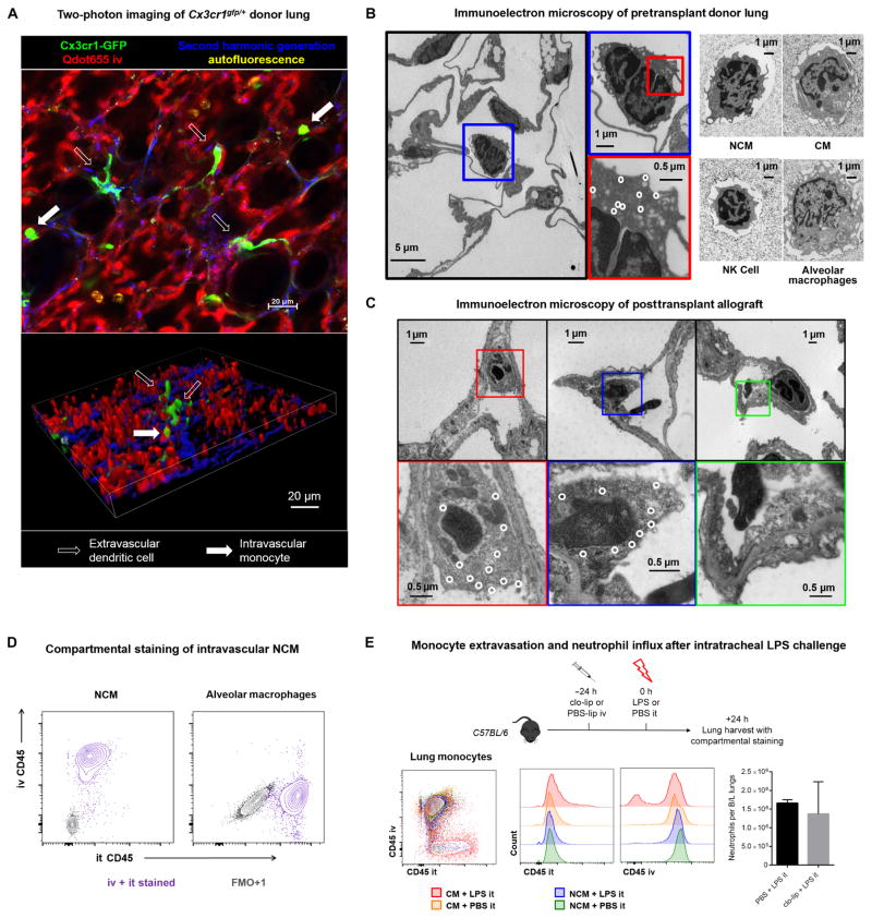
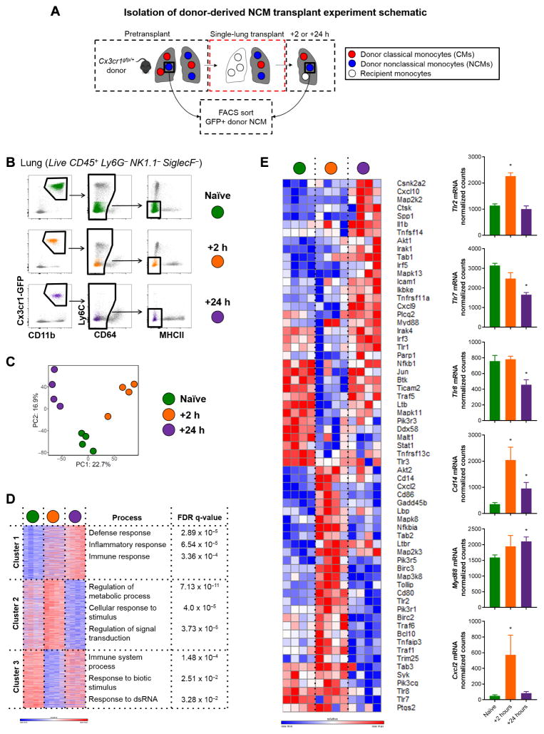
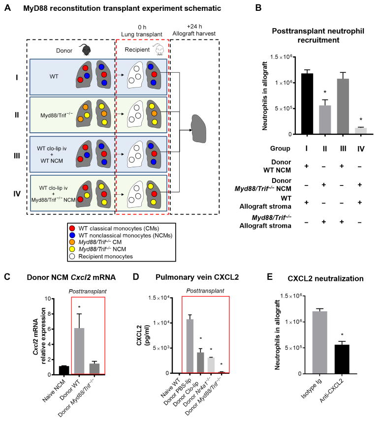
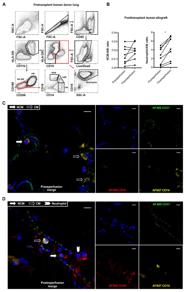
Primary graft dysfunction is the predominant driver of mortality and graft loss after lung transplantation. Recruitment of neutrophils as a result of ischemia-reperfusion injury is thought to cause primary graft dysfunction; however, the mechanisms that regulate neutrophil influx into the injured lung are incompletely understood. We found that donor-derived intravascular nonclassical monocytes (NCMs) are retained in human and murine donor lungs used in transplantation and can be visualized at sites of endothelial injury after reperfusion. When NCMs in the donor lungs were depleted, either pharmacologically or genetically, neutrophil influx and lung graft injury were attenuated in both allogeneic and syngeneic models. Similar protection was observed when the patrolling function of donor NCMs was impaired by deletion of the fractalkine receptor CX3CR1. Unbiased transcriptomic profiling revealed up-regulation of MyD88 pathway genes and a key neutrophil chemoattractant, CXCL2, in donor-derived NCMs after reperfusion. Reconstitution of NCM-depleted donor lungs with wild-type but not MyD88-deficient NCMs rescued neutrophil migration. Donor NCMs, through MyD88 signaling, were responsible for CXCL2 production in the allograft and neutralization of CXCL2 attenuated neutrophil influx. These findings suggest that therapies to deplete or inhibit NCMs in donor lung might ameliorate primary graft dysfunction with minimal toxicity to the recipient.


Conflict of interest statement

Competing interests: The authors declare that they have no competing interests.

Figures

Fig. 1
Fig. 1. Donor intravenous clo-lip treatment ameliorates PGD after transplantation
(A) Experimental design. (B) Allograft histology. Representative histology of the heart-lung blocks of control PBS-lip and intravenous (iv) clo-lip–treated donors at 24 hours after reperfusion. Hematoxylin and eosin staining. Inset scale bars, 50 μm. (C) Allograft function measured by PaO2 on 100% FiO2 as a marker of lung injury at 24 hours after transplant. *P = 0.01, n = 3 to 6 per group. (D) Allograft edema measured by wet-to-dry ratio of allograft at 4 and 24 hours after reperfusion. *P < 0.001, n = 5 per group. (E) Allograft vascular permeability measured by Evans blue dye extravasation leak test of allograft at 4 and 24 hours after reperfusion. *P < 0.01, n = 5 per group. Unpaired Student’s t test was used to compare means.
Fig. 2
Fig. 2. Intravenous clo-lip selectively depletes NCMs in donor lungs
(A) Experimental design. (B) Flow cytometry plots showing effects of monocyte depletion strategies on CMs, NCMs, and CD11b+ dendritic cells (DCs) in wild-type (WT) mice (full gating strategy is shown in fig. S2). (C) Effects of clo-lip and anti-CCR2 on the number of monocytes and relative composition of lung monocytes at 24 hours after treatment. *P < 0.01, n = 5 per group. (C) Data are representative of five experiments. Unpaired Student’s t test with Holm-Šidák correction for multiple comparisons was used to compare means.
Fig. 3
Fig. 3. Influx of recipient neutrophils into the allograft is abrogated by depletion of donor NCMs
(A) Experimental design. (B to G) Intravital two-photon imaging at 2 hours, starting at t = 0 min through t = 30 min after reperfusion. Representative still images of control PBS-lip–treated donor allograft immediately after reperfusion (also refer to movies S1 and S2). Green, LysM+; red, Qdot655 blood vessels. Unpaired Student’s t test was used to compare means. Scale bar, 50 μm. (H) Experimental design and result of differential monocyte depletion strategies in donors and recipients. Combinations of donor and recipient treatments were used to selectively deplete the different monocyte populations. The allograft was harvested at 24 hours after transplantation, and neutrophil influx was determined using flow cytometry. *P = 0.001 compared to group I (all other comparisons to group I are not significant), n = 6 per group. Unpaired Student’s t test with Holm-Šidák correction for multiple comparisons was used to compare means.
Fig. 4
Fig. 4. Pulmonary intravascular NCMs are dependent on CX3CR1 to recruit neutrophils, and reconstitution of depleted NCMs restores neutrophil influx in NR4A1-deficient mice
(A) Experimental design for CX3CR1 knockout transplants. (B) Representative flow plots and effects of CX3CR1 deletion on monocyte populations in donor lungs and posttransplant neutrophil infiltration of the allograft. *P = 0.01, n = 6 per group. Unpaired Student’s t test was used to compare means. (C) Experimental design for NR4A1 knockout transplants. (D) Representative flow plots and effects of NR4A1 deletion NCM reconstitution on monocyte populations in donor lungs and posttransplant neutrophil infiltration. *P = 0.01, n = 5 per group. Unpaired Student’s t test with Holm-Šidák correction for multiple comparisons was used to compare means.
Fig. 5
Fig. 5. Compartmentalization of pulmonary NCMs and CMs and their response to intratracheal lipopolysaccharide
(A) Two-photon imaging of Cx3cr1gfp/+ lungs. Green, Cx3cr1+; red, Qdot655 blood vessels; blue, second harmonic generation collagen; yellow, autofluorescent alveolar macrophages. Filled arrow, intravascular cell; open arrow, extravascular cell. (B) Morphology of the lung myeloid cell populations in the donor lung using immunogold electron microscopy in the Cx3cr1gfp/+ reporter mouse. Left: Immunoelectron microscopy of fixed Cx3cr1gfp/+ lung, with white circles highlighting gold nanoparticles staining for GFP. Right: Electron microscopy micrographs of postsort cells from WT B6 mouse lung. (C) Immunoelectron microscopy of Cx3cr1gfp/+ donor lungs at 4 hours after reperfusion, with white circles highlighting gold nanoparticles staining for GFP. Left and middle: NCMs bound to the endothelium with areas of exposed, thickened basement membrane and endothelial cell blebbing. Right: Neutrophil bound to the endothelium in the vicinity of a blebbing endothelial cell. (D) Representative compartmental staining of NCMs and alveolar macrophages. As negative control, FMO+1 (fluorescence minus one control + 1) staining is shown. it, intratracheal. (E) Experimental design and representative flow diagrams of intravenous and intratracheal anti-CD45 staining in LPS-treated and control mice along with neutrophil infiltration into the lungs with and without intravenous clo-lip pretreatment. Unpaired Student’s t test, not significant. n = 5 per group. LPS, lipopolysaccharide.
Fig. 6
Fig. 6. Transcriptional profiling of murine posttransplant donor-derived NCMs
(A) Experimental plan to isolate and sort NCMs from donor-naïve, +2-hour, and +24-hour posttransplant lung. (B) Sorting strategy with representative flow plots from each time point. (C) Principle components (PC) analysis of samples. (D) Gene ontology process enrichment analysis using K-means clustering. (E) TLR and nuclear factor κB signaling pathway genes, which were differentially expressed by cutoff of adjusted P value of <0.05 by pairwise comparison between time points, with the normalized counts of genes of interest depicted. *P < 0.04 by one-way analysis of variance (ANOVA), n = 4 per group. Heat map scale bars represent log2 scale.
Fig. 7
Fig. 7. NCMs are dependent on MyD88/TRIF signaling to produce CXCL2
(A) Experimental design. (B) Effects of reconstitution of NCM-depleted donor lungs with either WT or Myd88/Trif−/− NCMs. *P < 0.01, n = 5 per group. (C) Cxcl2 transcript expression level in donor-derived NCMs measured by quantitative polymerase chain reaction at 2 hours after transplant. As control, the baseline expression of Cxcl2 in WT pulmonary NCMs is shown. *P = 0.01. (D) Effect of NCM depletion on CXCL2 cytokine levels in allograft circulation when NCMs are depleted. As control, the CXCL2 levels in left pulmonary vein blood of WT mice are shown. *P < 0.01, n = 5 per group. Unpaired Student’s t test with Holm-Šidák correction for multiple comparisons was used to compare means. (E) Effect of CXCL2 blockade on neutrophil recruitment in the recipient at 24 hours after transplant. *P = 0.001, n = 5 per group. Unpaired Student’s t test was used to compare means.
Fig. 8
Fig. 8. Myeloid cell populations in human donor lungs and immediate postreperfusion changes
(A) Representative gating strategy of human lungs flushed and used in clinical transplantation. After excluding doublets and dead cells, and including only CD45+ cells, neutrophils were identified as CD15+CD16+SSChigh. After gating out CD15+ events, an HLA-DR+CD11b+ gate was used to identify monocytes and macrophages. Alveolar macrophages (AMs) were identified as CD15HLA-DR+CD11b+CD169+CD206+. After gating out AMs, NCMs were identified as CD16++CD14dim, intermediate monocytes (IntMs) as CD16+CD14+, and CMs as CD14+CD16. (B) Changes in NCMs and neutrophils at 90 min after reperfusion. Data are expressed as cell count per AM to standardize across patients. Biopsies were taken serially from the same location in the lung. *P = 0.02 (by paired Student’s t test), n = 8. Immunofluoresence microscopy of prereperfusion (C) and postreperfusion (D) human lung samples depicting endothelial-bound intravascular CD16+CD14dim NCMs (filled white arrow) in contrast with CD16 CD14high CMs (open arrow) and CD16+CD14+ neutrophils (filled white chevron). Green, CD31; blue, DAPI (4′,6-diamidino-2-phenylindole); red, CD16; yellow, CD14. Scale bars, 10 μm.

References

    1. Porteous MK, Diamond JM, Christie JD. Primary graft dysfunction: Lessons learned about the first 72 h after lung transplantation. Curr Opin Organ Transplant. 2015;20:506–514. - PMC - PubMed
    1. Christie JD, Carby M, Bag R, Corris P, Hertz M, Weill D ISHLT Working Group on Primary Lung Graft Dysfunction. Report of the ISHLT Working Group on Primary Lung Graft Dysfunction part II: Definition. A consensus statement of the International Society for Heart and Lung Transplantation. J Heart Lung Transpl. 2005;24:1454–1459. - PubMed
    1. Bharat A, Kuo E, Steward N, Aloush A, Hachem R, Trulock EP, Patterson GA, Meyers BF, Mohanakumar T. Immunological link between primary graft dysfunction and chronic lung allograft rejection. Ann Thorac Surg. 2008;86:189–195. - PMC - PubMed
    1. Daud SA, Yusen RD, Meyers BF, Chakinala MM, Walter MJ, Aloush AA, Patterson GA, Trulock EP, Hachem RR. Impact of immediate primary lung allograft dysfunction on bronchiolitis obliterans syndrome. Am J Respir Crit Care Med. 2007;175:507–513. - PubMed
    1. Belperio JA, Keane MP, Burdick MD, Londhe V, Xue YY, Li K, Phillips RJ, Strieter RM. Critical role for CXCR2 and CXCR2 ligands during the pathogenesis of ventilator-induced lung injury. J Clin Invest. 2002;110:1703–1716. - PMC - PubMed

Publication types