

Molecular Pathways: Targeting the Microenvironment of Liver Metastases
- PMID: 28615370
- PMCID: PMC5668192
- DOI: 10.1158/1078-0432.CCR-15-1636
Molecular Pathways: Targeting the Microenvironment of Liver Metastases
Abstract
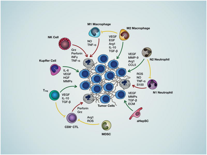
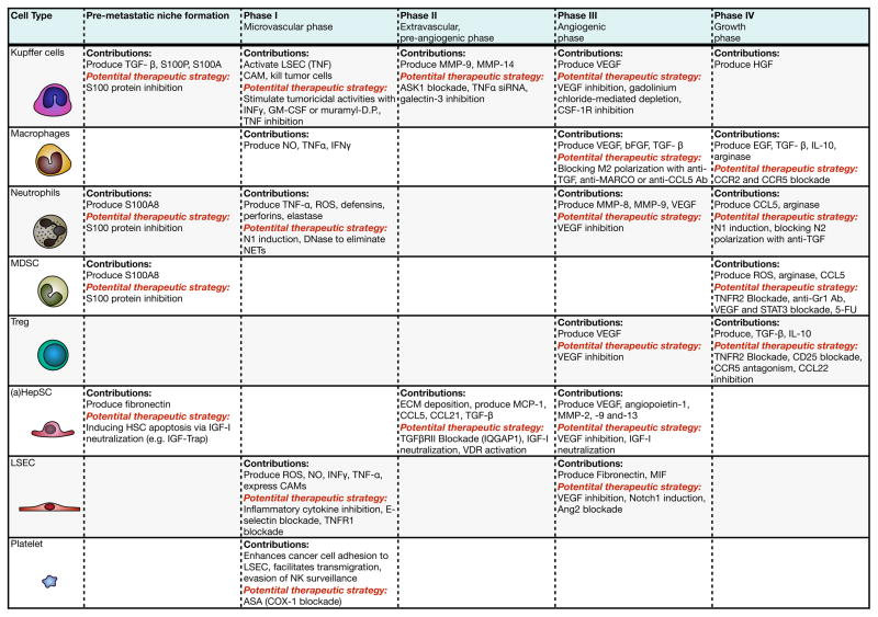
Curative treatment for metastatic solid cancers remains elusive. The liver, which is nourished by a rich blood supply from both the arterial and portal venous systems, is the most common site of visceral metastases, particularly from cancers arising in the gastrointestinal tract, with colorectal cancer being the predominant primary site in Western countries. A mounting body of evidence suggests that the liver microenvironment (LME) provides autocrine and paracrine signals originating from both parenchymal and nonparenchymal cells that collectively create both pre- and prometastatic niches for the development of hepatic metastases. These resident cells and their molecular mediators represent potential therapeutic targets for the prevention and/or treatment of liver metastases (LM). This review summarizes: (i) the current therapeutic options for treating LM, with a particular focus on colorectal cancer LM; (ii) the role of the LME in LM at each of its phases; (iii) potential targets in the LME identified through preclinical and clinical investigations; and (iv) potential therapeutic approaches for targeting elements of the LME before and/or after the onset of LM as the basis for future clinical trials. Clin Cancer Res; 23(21); 6390-9. ©2017 AACR.
©2017 American Association for Cancer Research.
Figures
References
-
- Turdean S, Gurzu S, Turcu M, Voidăzan S, Sin A. Liver Metastases: Incidence and Clinicopathological Data. Acta Medica Marisiensis. 2012;58(4):254–8.
-
- Chambers AF, Groom AC, MacDonald IC. Dissemination and growth of cancer cells in metastatic sites. Nat Rev Cancer. 2002;2(8):563–72. - PubMed
-
- Brodt P. Role of the Microenvironment in Liver Metastasis: From Pre- to Prometastatic Niches. Clin Cancer Res. 2016;22(24):5971–82. - PubMed
Publication types
MeSH terms
Grants and funding
LinkOut - more resources
Full Text Sources
Other Literature Sources
Medical
