. 2017 Jul 11;89(2):163-169.
doi: 10.1212/WNL.0000000000004089. Epub 2017 Jun 14.

Modulation of intrinsic resting-state fMRI networks in women with chronic migraine

Affiliations

Modulation of intrinsic resting-state fMRI networks in women with chronic migraine

X Michelle Androulakis et al. Neurology. .

Abstract

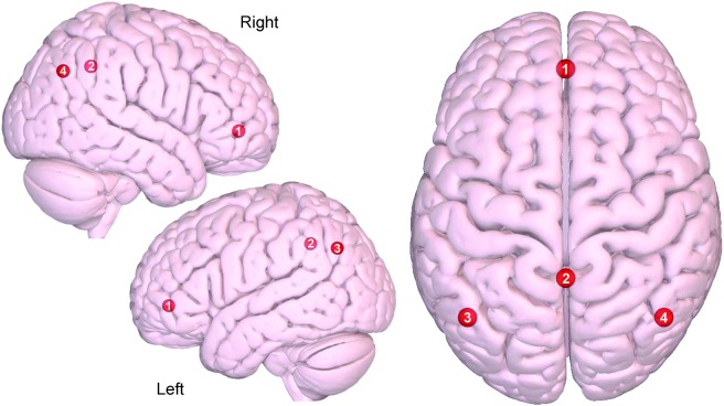
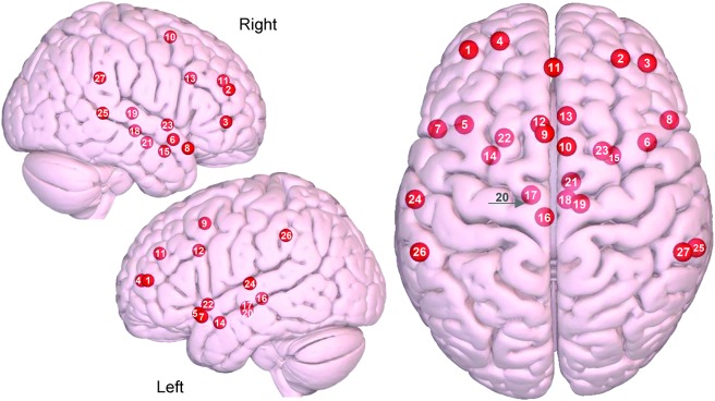
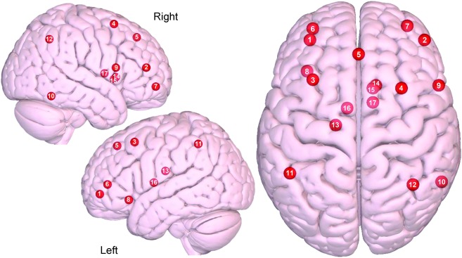
Objective: To evaluate the intrinsic resting functional connectivity of the default mode network (DMN), salience network (SN), and central executive network (CEN) network in women with chronic migraine (CM), and whether clinical features are associated with such abnormalities.

Methods: We analyzed resting-state connectivity in 29 women with CM as compared to age- and sex-matched controls. Relationships between clinical characteristics and changes in targeted networks connectivity were evaluated using a multivariate linear regression model.

Results: All 3 major intrinsic brain networks were less coherent in CM (DMN: p = 0.030, SN: p = 0.007, CEN: p = 0.002) as compared to controls. When stratified based on medication overuse headache (MOH) status, CM without MOH (DMN: p = 0.029, SN: p = 0.023, CEN: p = 0.003) and CM with MOH (DMN: p = 0.016, SN: p = 0.016, CEN: p = 0.015) were also less coherent as compared to controls. There was no difference in CM with MOH as compared to CM without MOH (DMN: p = 0.382, SN: p = 0.408, CEN: p = 0.419). The frequency of moderate and severe headache days was associated with decreased connectivity in SN (p = 0.003) and CEN (p = 0.015), while cutaneous allodynia was associated with increased connectivity in SN (p = 0.011).

Conclusions: Our results demonstrated decreased overall resting-state functional connectivity of the 3 major intrinsic brain networks in women with CM, and these patterns were associated with frequency of moderate to severe headache and cutaneous allodynia.


Figures

Figure 1
Figure 1. Axial and sagittal view of the default mode network
(1) Medial prefrontal, (2) precuneus/posterior cingulate cortex, (3) left lateral parietal, (4) right lateral parietal. Images were made with Surf Ice (nitrc.org/projects/surfice/) using the exact Montreal Neurologic Institute coordinate locations and sphere sizes. Node depth is illustrated by transparency.
Figure 2
Figure 2. Axial and sagittal view of the salience network
(1) Left dorsolateral prefrontal cortex (DLPFC), (2) right DLPFC, (3) right ventrolateral prefrontal cortex, (4) left frontal pole, (5) left orbital frontal insula, (6) right orbital frontal insula, (7) left temporal pole, (8) right temporal pole, (9) left supplementary motor area (SMA)/pre-SMA, (10) right SMA/pre SMA, (11) paracingulate cortex, (12) left dorsal anterior cingulate cortex (dACC), (13) right dACC, (14) left sublenticular extended amygdala, (15) right sublenticular extended amygdala, (16) left periaqueductal gray, (17) left hypothalamus, (18) right hypothalamus, (19) right dorsomedial thalamus, (20) left substantia nigra/ventral tegmental area, (21) right substantia nigra/ventral tegmental area, (22) left ventral striatum/pallidum, (23) right ventral striatum/pallidum, (24) left superior temporal, (25) right superior temporal, (26) left supramarginal gyrus (SMG), (27) right SMG. Images were made with Surf Ice (nitrc.org/projects/surfice/) using the exact Montreal Neurologic Institute coordinate locations. Node depth is illustrated by transparency.
Figure 3
Figure 3. Axial and sagittal view of the central executive network
(1) Left dorsolateral prefrontal cortex (DLPFC), (2) right DLPFC, (3) left DLPFC/frontal eye fields (FEF), (4) right DLPFC/FEF, (5) dorsal medial prefrontal cortex, (6) left ventrolateral prefrontal cortex (VLPFC), (7) right VLPFC, (8) left orbital frontal insula, (9) right inferior frontal gyrus, (10) right inferior temporal, (11) left lateral parietal, (12) right lateral parietal, (13) left dorsal caudate, (14) right dorsal caudate, (15) right ventromedial caudate, (16) left anterior thalamus, (17) right anterior thalamus. Images were made with Surf Ice (nitrc.org/projects/surfice/) using the exact Montreal Neurologic Institute coordinate locations. Node depth is illustrated by transparency.

References

    1. May A, Schulte LH. Chronic migraine: risk factors, mechanisms and treatment. Nat Rev Neurosci 2016;12:455–464. - PubMed
    1. Maizels M, Aurora S, Heinricher M. Beyond neurovascular: migraine as a dysfunctional neurolimbic pain network. Headache 2012;52:1553–1565. - PubMed
    1. Schwedt TJ, Chiang CC, Chong CD, Dodick DW. Functional MRI of migraine. Lancet Neurol 2015;14:81–91. - PMC - PubMed
    1. Pizoli CE, Shah MN, Snyder AZ, et al. . Resting-state activity in development and maintenance of normal brain function. Proc Natl Acad Sci USA 2011;108:11638–11643. - PMC - PubMed
    1. Foster BL, He BJ, Honey CJ, Jerbi K, Maier A, Saalmann YB. Spontaneous neural dynamics and multi-scale network organization. Front Syst Neurosci 2016;10:7. - PMC - PubMed