. 2017 Jul 28;292(30):12653-12666.
doi: 10.1074/jbc.M117.793018. Epub 2017 Jun 14.

Deconvoluting AMP-activated protein kinase (AMPK) adenine nucleotide binding and sensing

Affiliations

Deconvoluting AMP-activated protein kinase (AMPK) adenine nucleotide binding and sensing

Xin Gu et al. J Biol Chem. .

Abstract

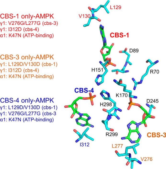
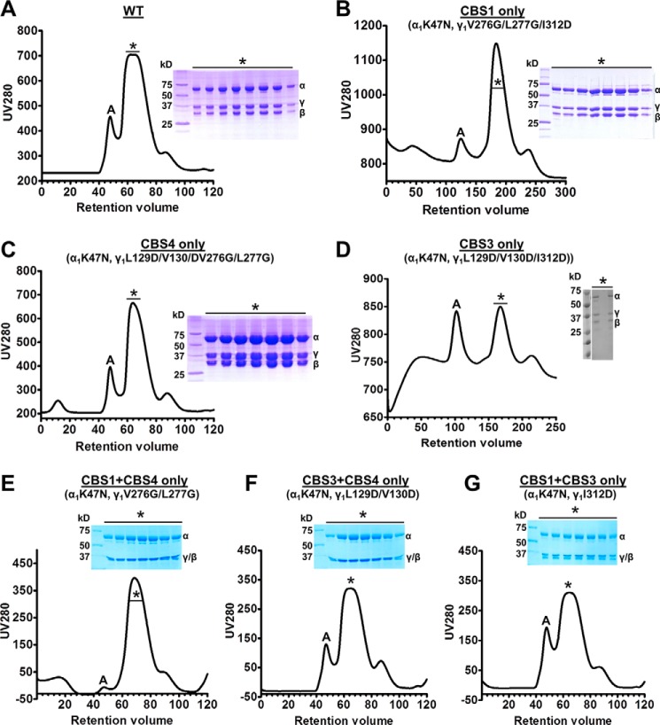
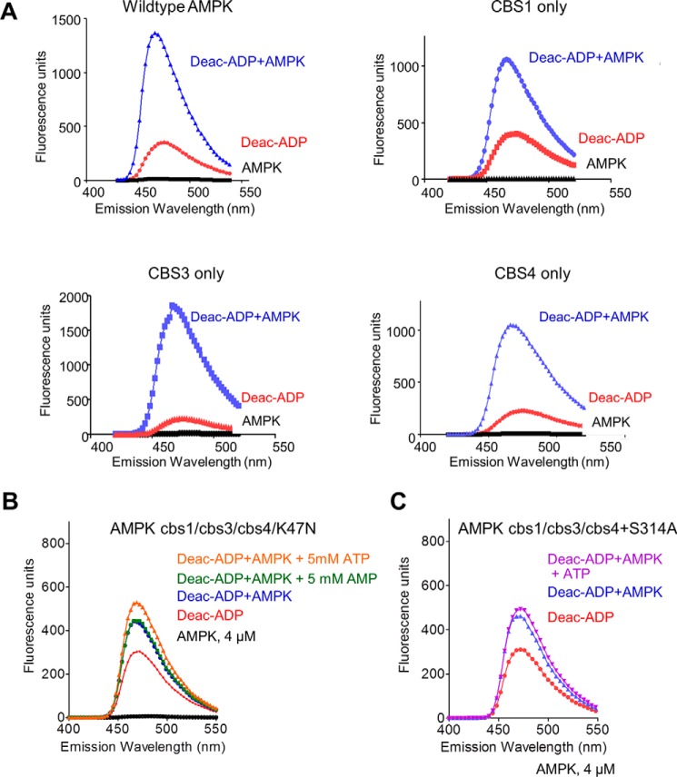
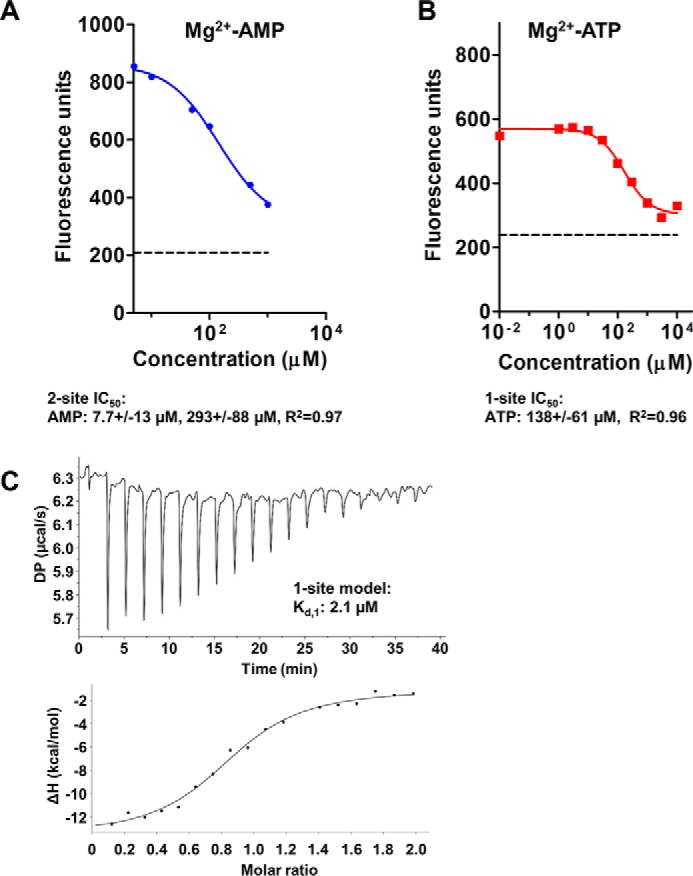
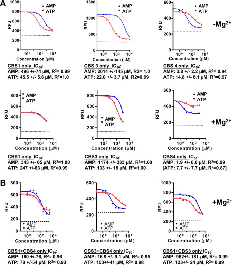
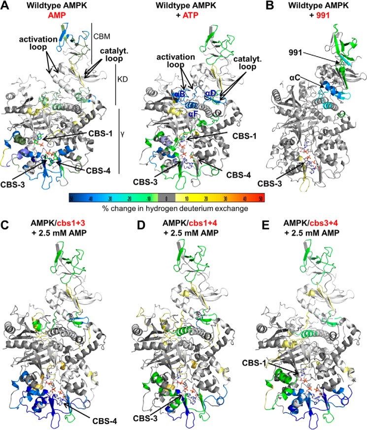
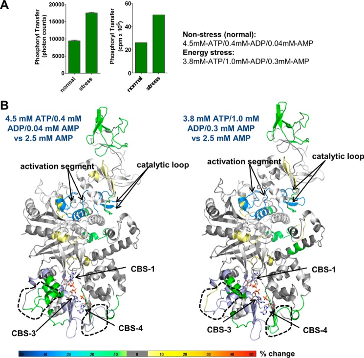
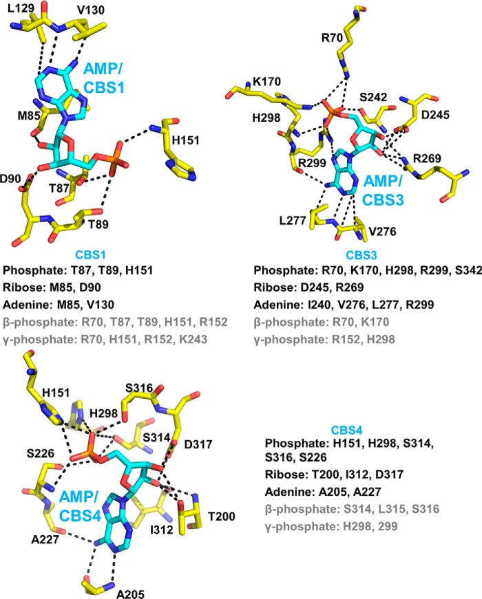
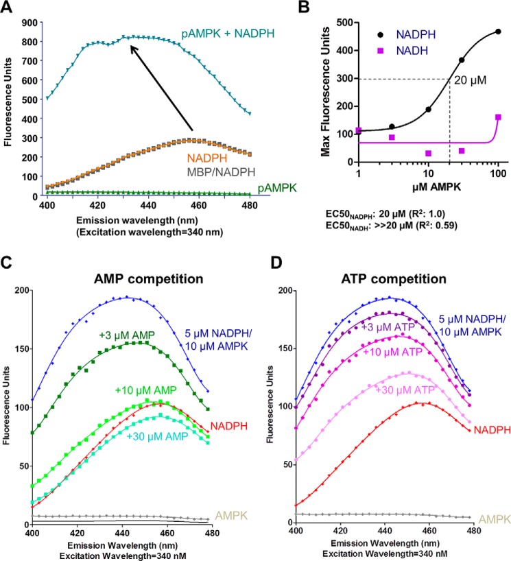
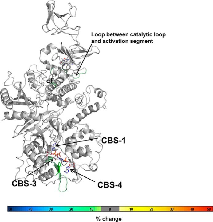
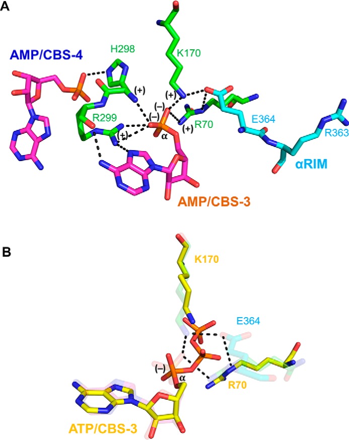
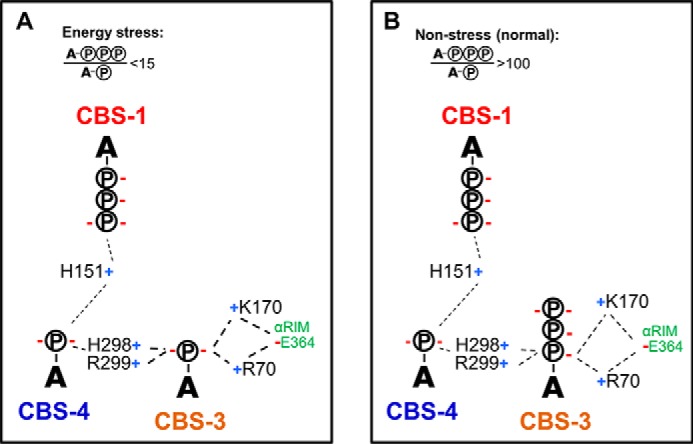
AMP-activated protein kinase (AMPK) is a central cellular energy sensor that adapts metabolism and growth to the energy state of the cell. AMPK senses the ratio of adenine nucleotides (adenylate energy charge) by competitive binding of AMP, ADP, and ATP to three sites (CBS1, CBS3, and CBS4) in its γ-subunit. Because these three binding sites are functionally interconnected, it remains unclear how nucleotides bind to individual sites, which nucleotides occupy each site under physiological conditions, and how binding to one site affects binding to the other sites. Here, we comprehensively analyze nucleotide binding to wild-type and mutant AMPK protein complexes by quantitative competition assays and by hydrogen-deuterium exchange MS. We also demonstrate that NADPH, in addition to the known AMPK ligand NADH, directly and competitively binds AMPK at the AMP-sensing CBS3 site. Our findings reveal how AMP binding to one site affects the conformation and adenine nucleotide binding at the other two sites and establish CBS3, and not CBS1, as the high affinity exchangeable AMP/ADP/ATP-binding site. We further show that AMP binding at CBS4 increases AMP binding at CBS3 by 2 orders of magnitude and reverses the AMP/ATP preference of CBS3. Together, these results illustrate how the three CBS sites collaborate to enable highly sensitive detection of cellular energy states to maintain the tight ATP homeostastis required for cellular metabolism.

Keywords: AMP; AMP-activated kinase (AMPK); ATP; CBS; HDX-MS; NADPH; energy metabolism; protein kinase.


Conflict of interest statement

The authors declare that they have no conflicts of interest with the contents of this article

Figures

Figure 1.
Figure 1.
Binding of AMP to the γ1 subunit. Stick presentation of the three AMP molecules (green carbon atoms) and key AMP-binding residues of AMPK (cyan carbon atoms). Oxygen is colored in red, nitrogen in blue, and phosphorus in orange. The phosphate groups of the 3 AMP molecules are coordinately bound by a set of charged amino acids, whereas the ribose and adenine rings face away from each other and are bound by CBS-specific residues. Mutations of the indicated adenine-binding residues therefore selectively block AMP binding to individual CBS sites. Note that the adenine-binding mutations at CBS3 and CBS4 are each sufficient, even at the high concentration of adenine nucleotides in cells, to completely abolish AMPK activation by AMP without affecting AMPK catalytic activity in the absence of AMP (18).
Figure 2.
Figure 2.
Size exclusion chromatograms and SDS-PAGE of AMPK mutant proteins. A, C, and E–G, 120-ml column; B and D, 300-ml column; *, monomeric α1β2γ1 fractions; A, aggregate/oligomers.
Figure 3.
Figure 3.
Fluorescence spectra of wild-type and mutant AMPK/deac-ADP and competition by AMP and ATP. A, emission spectra of 0.5 μm deac-ADP in the absence and presence of wild-type and mutant AMPK proteins in which only one of the CBS sites remained functional (CBS1 only: α1-K47N, γ1-V276G/L277G/I312D; CBS3 only: α1-K47N, γ1-L129D/V130D/I312D; CBS4 only: α1-K47N, γ1-L129D/V130D/V276G/L277G). Excitation: 430 nm. B and C, emission spectra of 0.5 μm deac-ADP in the absence and presence of 4 μm α1β2γ1 AMPK (α1-K47N, γ1-L129D/V130D/V276G/L277G/I312D) (B) or α1β2γ1 AMPK (α1-K47N, γ1-L129D/V130D/V276G/L277G/I312D/S314A) (C) and 5 mm AMP or ATP.
Figure 4.
Figure 4.
Nucleotide binding to wild-type AMPK. A and B, competition of 0.5 μm deac-ADP bound to α1β2γ1 AMPK by unlabeled Mg2+-AMP (A) and Mg2+-ATP (B). The dashed line indicates the fluorescence signal of unbound deac-ADP. Because fluorescence competition failed to unambiguously determine the IC50 value for the high affinity AMP-binding site, we independently determined its affinity by ITC (C).
Figure 5.
Figure 5.
Nucleotide binding to individual CBSs and pairs of CBSs. Competition of 0.5 μm deac-ADP bound to 4 μm α1β2γ1 AMPK by unlabeled AMP and ATP. A, AMPK mutants with only a single functional adenine nucleotide-binding site (see Fig. 1). Note that in these experiments singly and doubly mutated CBS4 (I312D and I312D/S314A) behaved identically. The ability of Mg2+-AMP to compete deac-ADP from 4 μm AMPK with an IC50 of 1.9 μm indicates that AMP still binds CBS4 with submicromolar affinity, but no longer non-exchangeably. B, AMPK mutants with two functional adenine nucleotide-binding sites. Samples were incubated for 30 min, excited at 430 nm, and emission recorded at 470 nm. The dashed line indicates the fluorescence signal of unbound (i.e. completely competed) deac-ADP.
Figure 6.
Figure 6.
HDX-MS changes of wild-type and mutant AMPK upon incubation with AMP or ATP. Changes were overlaid as heat map onto the structure of α1β2γ1 AMPK (4RER). The heat map legend (% change) is shown at the bottom. A, AMPK wild-type in the presence versus absence of AMP (left) or ATP (right). B, AMPK wild-type in the presence versus absence of 991. C, D, and E, AMPK triple mutants in which only CBS4 (C), CBS3 (B), or CBS1 (E) are functional (see Fig. 1).
Figure 7.
Figure 7.
Changes in HDX-MS protection in the presence of AXP mixtures mimicking non-stress and energy stress conditions. A, AMPK kinase assay in the presence of AXP mixtures mimicking nonstress (4.5 mm ATP, 0.4 mm ADP, 0.04 mm AMP) and energy stress (3.8 mm ATP, 1.0 mm ADP, 0.3 mm AMP) conditions. Left panel, AlphaScreen-based kinase assay; right panel, radioactive kinase assay. B, HDX-MS perturbation map of AMPK in the presence of the two different AXP mixtures. HDX-MS changes were overlaid as heat map onto the structure of α1β2γ1 AMPK (PDB code 4RER). Main areas of differential protection are highlighted by dashed outlines. Note that there is no change at the catalytic site, indicating that the catalytic site is constitutively bound by ATP as expected. The color code (% change) is shown below the structures. The relatively mild changes are consistent with noticeable ATP exchange only at CBS3, whereas the non-physiological shift from high concentration of pure AMP to high concentration of pure ATP resulted in strong HDX changes (Fig. 6A), consistent with near full nucleotide exchange at both CBS3, CBS1, and ATP binding/release at the catalytic site.
Figure 8.
Figure 8.
AXP/CBS interaction network in human AMPK-γ1. Shown are the three AMP-bound CBS sites in holo-AMPK co-crystallized with AMP. β- and γ-phosphate interacting residues (gray) are collectively based on the structures of core AMPK co-crystallized with ATP (PDB code 4EAG) (20) and core- AMPK soaked with ATP (PDB codes 2V92 and 4EAJ) (19, 20).
Figure 9.
Figure 9.
NADPH binds adenine nucleotide-binding site(s) in the γ-subunit. A, NADPH emission spectra in the absence and presence of MBP and MBP-tagged phosphorylated α1β2γ1 AMPK. The spectrum of AMPK in the absence is shown as negative control. B, estimation of NADPH and NADH binding constants. 5 μm NADPH or NADH were incubated with increasing concentrations of MBP-AMPK and MBP-AMPK concentrations were plotted against fluorescence intensity at the emission maxima. C and D, AMP and ATP compete the NADPH binding signal. Emission spectra of 5 μm NADPH, 10 μm AMPK were recorded in the absence and presence of increasing concentrations of AMP (C) and ATP (D).
Figure 10.
Figure 10.
Binding of NADPH protects CBS3 and CBS4. HDX-MS changes wild-type AMPK upon incubation with 1 mm NADPH. Changes were overlaid as heat map onto the structure of α1β2γ1 AMPK (PDB code 4RER). The heat map legend (% change) is shown at the bottom.
Figure 11.
Figure 11.
CBS4 and αRIM stabilize AMP-binding at CBS3 and increase the AMP/ATP-binding preference. A, CBS4-CBS3-αRIM network in AMP-bound AMPK (PDB code 4CFE). B, transparent structure from panel A overlaid with ATP, Arg70, and Lys170 from the structure of ATP-soaked core-AMPK (PDB code 2V92). Note that the α-phosphate of ATP has only a single negative charge and that the β- and γ-phosphate groups of ATP push Lys170 and Arg70 away from αRIM Glu364.
Figure 12.
Figure 12.
Schematic model of AXP occupancy under energy-stress (A) and non-stress (B) conditions. Positive and negative charges are indicated by blue “+” and red “−” signs. A-circled P, AMP; A-3 circled P, ATP.

References

    1. Bullon P., Marin-Aguilar F., and Roman-Malo L. (2016) AMPK/mitochondria in metabolic diseases. EXS 107, 129–152 - PubMed
    1. Cheng J., Zhang T., Ji H., Tao K., Guo J., and Wei W. (2016) Functional characterization of AMP-activated protein kinase signaling in tumorigenesis. Biochim. Biophys. Acta 1866, 232–251 - PMC - PubMed
    1. Hardie D. G., Ross F. A., and Hawley S. A. (2012) AMP-activated protein kinase: a target for drugs both ancient and modern. Chem. Biol. 19, 1222–1236 - PMC - PubMed
    1. Mihaylova M. M., and Shaw R. J. (2011) The AMPK signalling pathway coordinates cell growth, autophagy and metabolism. Nat. Cell. Biol. 13, 1016–1023 - PMC - PubMed
    1. Steinberg G. R., and Kemp B. E. (2009) AMPK in health and disease. Physiol. Rev. 89, 1025–1078 - PubMed

Publication types