. 2017 Jun 14;7(1):3501.
doi: 10.1038/s41598-017-03256-0.

Transcriptome analysis reveals similarities between human blood CD3- CD56bright cells and mouse CD127+ innate lymphoid cells

Affiliations

Transcriptome analysis reveals similarities between human blood CD3- CD56bright cells and mouse CD127+ innate lymphoid cells

David S J Allan et al. Sci Rep. .

Abstract

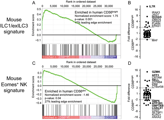
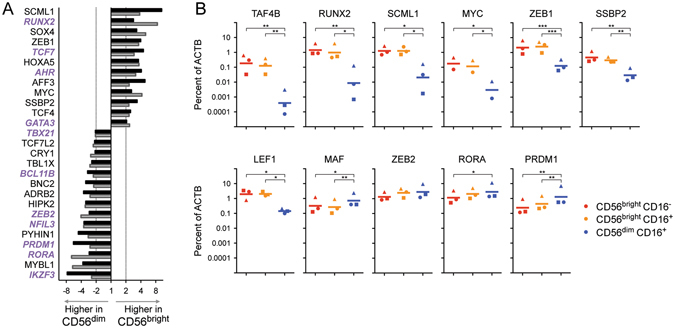
For many years, human peripheral blood natural killer (NK) cells have been divided into functionally distinct CD3- CD56bright CD16- and CD3- CD56dim CD16+ subsets. Recently, several groups of innate lymphoid cells (ILC), distinct from NK cells in development and function, have been defined in mouse. A signature of genes present in mouse ILC except NK cells, defined by Immunological Genome Project studies, is significantly over-represented in human CD56bright cells, by gene set enrichment analysis. Conversely, the signature genes of mouse NK cells are enriched in human CD56dim cells. Correlations are based upon large differences in expression of a few key genes. CD56bright cells show preferential expression of ILC-associated IL7R (CD127), TNFSF10 (TRAIL), KIT (CD117), IL2RA (CD25), CD27, CXCR3, DPP4 (CD26), GPR183, and MHC class II transcripts and proteins. This could indicate an ontological relationship between human CD56bright cells and mouse CD127+ ILC, or conserved networks of transcriptional regulation. In line with the latter hypothesis, among transcription factors known to impact ILC or NK cell development, GATA3, TCF7 (TCF-1), AHR, SOX4, RUNX2, and ZEB1 transcript levels are higher in CD56bright cells, while IKZF3 (AIOLOS), TBX21 (T-bet), NFIL3 (E4BP4), ZEB2, PRDM1 (BLIMP1), and RORA mRNA levels are higher in CD56dim cells.


Conflict of interest statement

The authors declare that they have no competing interests.

Figures

Figure 1
Figure 1
Signature genes, higher in mouse ILC subsets, are enriched in human blood CD3 CD56bright cells, while mouse NK cell signature genes are enriched in human CD3 CD56dim cells. (A,B) Transcriptional signatures of mouse NK cells (Core NK) and other mouse ILC (Core ILC) were defined by Robinette and colleagues as part of the ImmGen project. Enrichment of corresponding homologous human genes was examined by Gene Set Enrichment Analysis in human blood CD56bright CD16 CD3 and CD56dim CD16+ CD3 populations characterized by Affymetrix HTA2.0 microarray. Deviations of cumulative scores (green line) above zero indicate enrichment in CD56bright cells, while negative scores indicate enrichment in CD56dim cells. Samples of each subset from three individuals were used for analysis. Signature genes without human homologues were not included in the analysis. (C) Flow cytometry staining of gated human blood CD56bright CD16 CD3 or CD56dim CD16+ CD3 populations from three donors. Quadrant gates were set using fluorescence minus one (FMO) stains, of which one is shown. Intracellular staining for EOMES was performed on three blood samples distinct from those used to acquire the other results.
Figure 2
Figure 2
Expression differences between mouse splenic CD127+ NK1.1+ NKp46+ ILC and CD127 NK1.1+ NKp46+ NK cells show commonalities with differences between human blood CD56bright and CD56dim cells. ImmGen microarray data examining mouse splenic NK1.1+ NKp46+ CD127+ CD27+ cells (annotated as ILC1) and NK1.1+ NKp46+ CD127 CD27+/− NK cells is analysed to show fold-change difference between subsets (x-axis). For all mouse genes with corresponding human genes in the NCBI Homologene database, this is contrasted with microarray expression differences observed between human blood CD56bright and CD56dim populations (y-axis) (HTA2.0 microarray). Transcripts differing by ≥2-fold between both human and mouse subsets are labelled, and their numbers are shown. Transcripts similarly varying by ≥2-fold in mouse ILC1/NK comparisons in both liver and small intestine (in addition to spleen) are shown in blue; those differing by ≥2-fold in 2-of-3 comparisons of mouse tissues are in green (see Suppl. Figure 3). Bold names also show ≥2-fold differences in an independent microarray dataset comparing human CD56bright and CD56dim cells (see Suppl. Figure 4). Although not matched by Homologene, human HLA-DRA (paired with mouse H2-Aa) is shown (with asterisk) but not included in transcript counts. (B) Flow cytometry staining of gated human blood CD56bright CD16 CD3 or CD56dim CD16+ CD3 populations from three donors. Quadrant gates were set using fluorescence minus one (FMO) stains.
Figure 3
Figure 3
Transcripts with higher expression in mouse ILC1 and ex-ILC3 subsets show enrichment in human CD56bright cells. Lists of signature transcripts were compiled from published microarray data comparing three mouse NK1.1+ NKp46+ subsets: (i) Eomes+ NK cells, (ii) Rorγtfate map+ ex-ILC3 (that formerly expressed Rorγt), and (iii) ILC1 that expressed neither (from ref. 12). Enrichment of corresponding homologous human genes was examined by Gene Set Enrichment Analysis in human CD56bright and CD56dim populations characterized by Affymetrix HTA2.0 microarray, using the following signatures: (A-B) Mouse ILC1/ex-ILC3 signature (transcripts detected at ≥2-fold higher levels in both ILC1 versus NK and ex-ILC3 versus NK comparisons and (C-D) Mouse Eomes +NK cell signature (genes with ≥2-fold higher expression in both NK versus ILC1 and NK versus ex-ILC3 comparisons). (B,D) Fold difference in expression of these transcripts between human CD56bright and CD56dim cells is shown for comparison. Genes differing by ≥2-fold between the human subsets are labelled (in order by fold change difference) and emboldened if conserved in similar analyses with a second human microarray dataset (see Suppl. Figure 5).
Figure 4
Figure 4
Human CD56bright and CD56dim subsets differ in expression of many transcription factors known to affect development of ILC and NK cells. (A) Fold-change differences in expression between CD56bright and CD56dim cells analysed by two Affymetrix microarray platforms: HTA2.021 (black bars) and HG-U133 A/B22 (grey bars). Three samples of each subset were analysed on each microarray, for a total of 6 independent sample pairs. Transcription factors showing ≥2-fold difference on both array platforms are shown, ordered by mean fold change. Genes labelled in purple italics have been shown to affect development of ILC and/or NK in mouse models (see references in text). (B) Quantitative real-time RT-PCR validation of several differences in transcription factor expression (including some that showed ≥2-fold change in only one microarray dataset). RNA was isolated from human blood CD56bright CD16 cells, CD56dim CD16+ cells, and the “intermediate population” displaying CD56bright CD16+ phenotype. Transcript levels are quantitated relative to ACTB in samples from three individuals (represented by triangle, circle and square symbols) with a bar depicting the geometric mean level of expression. Samples were distinct from those analysed by microarray. *p < 0.05, **p < 0.01, ***p < 0.001, by Tukey’s Multiple Comparison Test after one way ANOVA of Log10 transformed values. Trends were consistent if data was normalized to GAPDH transcript levels instead of ACTB.

References

    1. Lanier LL, Le AM, Civin CI, Loken MR, Phillips JH. The relationship of CD16 (Leu-11) and Leu-19 (NKH-1) antigen expression on human peripheral blood NK cells and cytotoxic T lymphocytes. J Immunol. 1986;136:4480–4486. - PubMed
    1. Cooper MA, Fehniger TA, Caligiuri MA. The biology of human natural killer-cell subsets. Trends Immunol. 2001;22:633–640. doi: 10.1016/S1471-4906(01)02060-9. - DOI - PubMed
    1. Cooper MA, et al. Human natural killer cells: a unique innate immunoregulatory role for the CD56(bright) subset. Blood. 2001;97:3146–3151. doi: 10.1182/blood.V97.10.3146. - DOI - PubMed
    1. Chan A, et al. CD56bright human NK cells differentiate into CD56dim cells: role of contact with peripheral fibroblasts. J Immunol. 2007;179:89–94. doi: 10.4049/jimmunol.179.1.89. - DOI - PubMed
    1. Huntington ND, et al. IL-15 trans-presentation promotes human NK cell development and differentiation in vivo. J Exp Med. 2009;206:25–34. doi: 10.1084/jem.20082013. - DOI - PMC - PubMed

Publication types