

An Anatomical study for localisation of Zygomatic branch of Facial nerve and Masseteric nerve - An aid to nerve coaptation for facial reanimation surgery: A cadaver based study in Eastern India
- PMID: 28615814
- PMCID: PMC5469240
- DOI: 10.4103/ijps.IJPS_128_16
An Anatomical study for localisation of Zygomatic branch of Facial nerve and Masseteric nerve - An aid to nerve coaptation for facial reanimation surgery: A cadaver based study in Eastern India
Abstract
Context: In cases of chronic facial palsy, where direct neurotisation is possible, ipsilateral masseteric nerve is a very suitable motor donor. We have tried to specifically locate the masseteric nerve for this purpose.
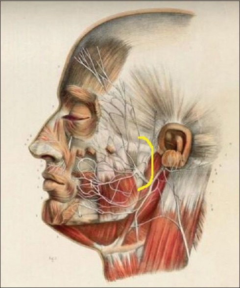
Aims: Describing an approach of localisation and exposure of both the zygomatic branch of Facial nerve and the nerve to masseter, with respect to a soft tissue reference point over face.
Settings and design: Observational cross sectional study, conducted on 12 fresh cadavers.
Subjects and methods: A curved incision was given, passing about 0.5cms in front of the tragal cartilage. A reference point "R" was pointed out. The zygomatic branch of facial nerve and masseteric nerve were dissected out and their specific locations were recorded from fixed reference points with help of copper wire and slide callipers.
Statistical analysis used: Central Tendency measurements and Unpaired "t" test.
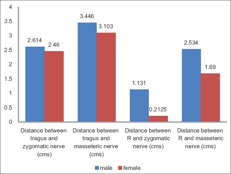
Results: Zygomatic branch of the Facial nerve was located within a small circular area of radius 1 cm, the centre of which lies at a distance of 1.1 cms (±0.4cm) in males and 0.2cm (±0.1cm) in females from the point, 'R', in a vertical (coronal) plane. The nerve to masseter was noted to lie within a circular area of 1 cm radius, the centre of which was at a distance of 2.5cms (±0.4cm) and 1.7cms (±0.2cm) from R, in male and female cadavers, respectively. Finally, Masseteric nerve's depth, from the masseteric surface was found to be 1cm (±0.1cm; male) and 0.8cm (±0.1cm; female).
Conclusions: This novel approach can reduce the post operative cosmetic morbidity and per-operative complications of facial reanimation surgery.
Keywords: Chronic facial palsy; masseteric nerve; zygomatic branch (of facial nerve).
Conflict of interest statement
There are no conflicts of interest.
Figures


References
-
- Javeed Ahmad S, Hamid Rather A. Prospective study of physical therapy in facial nerve paralysis: Experience at a multispeciality hospital of Kashmir. J Med Sci. 2012;15:145–8.
-
- O’Brien BM, Franklin JD, Morrison WA. Cross-facial nerve grafts and microneurovascular free muscle transfer for long established facial palsy. Br J Plast Surg. 1980;33:202–15. - PubMed
-
- Terzis JK, Konofaos P. Nerve transfers in facial palsy. Facial Plast Surg. 2008;24:177–93. - PubMed
LinkOut - more resources
Full Text Sources
Other Literature Sources