. 2017 Jun 12:9:6.
doi: 10.1186/s11689-017-9184-y. eCollection 2017.

Acamprosate in a mouse model of fragile X syndrome: modulation of spontaneous cortical activity, ERK1/2 activation, locomotor behavior, and anxiety

Affiliations

Acamprosate in a mouse model of fragile X syndrome: modulation of spontaneous cortical activity, ERK1/2 activation, locomotor behavior, and anxiety

Tori L Schaefer et al. J Neurodev Disord. .

Abstract

Background: Fragile X Syndrome (FXS) occurs as a result of a silenced fragile X mental retardation 1 gene (FMR1) and subsequent loss of fragile X mental retardation protein (FMRP) expression. Loss of FMRP alters excitatory/inhibitory signaling balance, leading to increased neuronal hyperexcitability and altered behavior. Acamprosate (the calcium salt of N-acetylhomotaurinate), a drug FDA-approved for relapse prevention in the treatment of alcohol dependence in adults, is a novel agent with multiple mechanisms that may be beneficial for people with FXS. There are questions regarding the neuroactive effects of acamprosate and the significance of the molecule's calcium moiety. Therefore, the electrophysiological, cellular, molecular, and behavioral effects of acamprosate were assessed in the Fmr1-/y (knock out; KO) mouse model of FXS controlling for the calcium salt in several experiments.

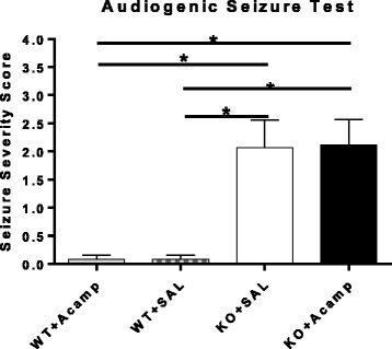
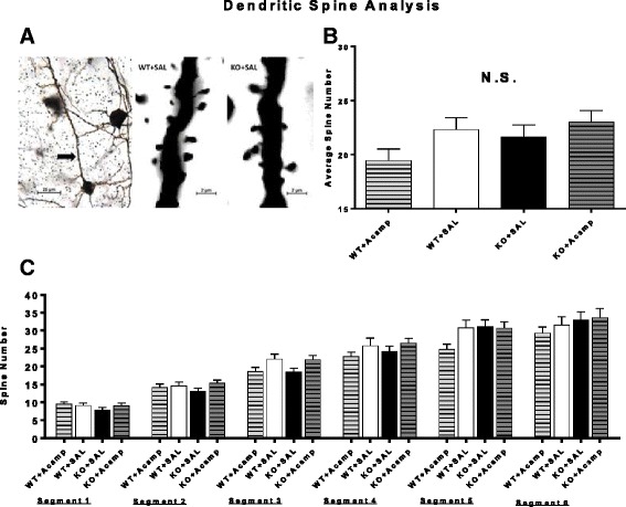
Methods: Fmr1 KO mice and their wild-type (WT) littermates were utilized to assess acamprosate treatment on cortical UP state parameters, dendritic spine density, and seizure susceptibility. Brain extracellular-signal regulated kinase 1/2 (ERK1/2) activation was used to investigate this signaling molecule as a potential biomarker for treatment response. Additional adult mice were used to assess chronic acamprosate treatment and any potential effects of the calcium moiety using CaCl2 treatment on behavior and nuclear ERK1/2 activation.

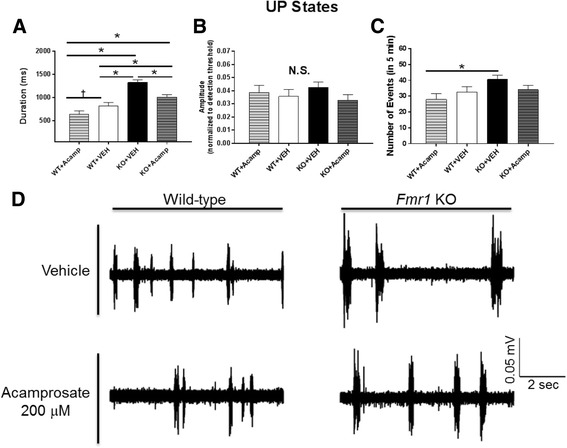
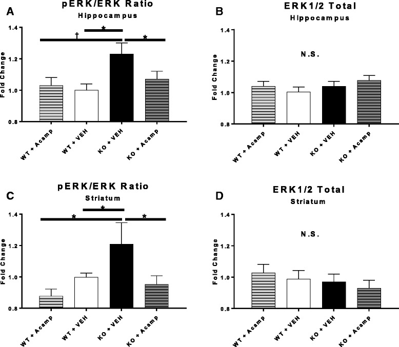
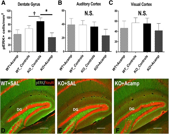
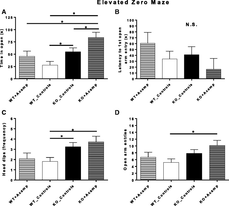
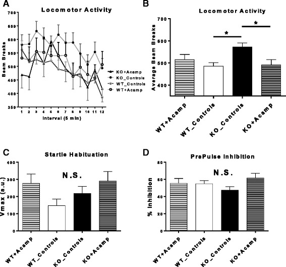
Results: Acamprosate attenuated prolonged cortical UP state duration, decreased elevated ERK1/2 activation in brain tissue, and reduced nuclear ERK1/2 activation in the dentate gyrus in KO mice. Acamprosate treatment modified behavior in anxiety and locomotor tests in Fmr1 KO mice in which control-treated KO mice were shown to deviate from control-treated WT mice. Mice treated with CaCl2 were not different from saline-treated mice in the adult behavior battery or nuclear ERK1/2 activation.

Conclusions: These data indicate that acamprosate, and not calcium, improves function reminiscent of reduced anxiety-like behavior and hyperactivity in Fmr1 KO mice and that acamprosate attenuates select electrophysiological and molecular dysregulation that may play a role in the pathophysiology of FXS. Differences between control-treated KO and WT mice were not evident in a recognition memory test or in examination of acoustic startle response/prepulse inhibition which impeded conclusions from being made about the treatment effects of acamprosate in these instances.

Keywords: Anxiety; Dendritic spine density; Electrophysiology; Extracellular signal-related kinase; Fragile X syndrome; Hippocampus; Hyperactivity; Open field; Striatum.


Figures

Fig. 1
Fig. 1
UP state recordings. Spontaneous UP states were measured in slices from P18–25 mice for 5 min in layer IV of the somatosensory cortex. Duration (a), amplitude (b), and number of events (c) were analyzed by two-way ANOVA with pairwise comparisons corrected using FDR method (two-tailed). Representative traces are shown in panel (d). There was a significant increase in UP state duration in the KO + VEH-treated mice compared to the WT + VEH-treated mice indicating a baseline effect of genotype. Bath application of 200 μM acamprosate significantly decreased the elevated UP state duration in the KO mice indicating a significant treatment although the acamprosate-treated KO slices still had UP state durations that were longer than WT + VEH slices. There was a trend towards a decreased UP state duration in the WT + Acamp group compared to the WT + VEH group. For number of events, there was a main effect of gene, and the KO + VEH slices had more UP state events than the WT + Acamp-treated mice. No change in amplitude was observed. WT + VEH, n = 16; WT + Acamp, n = 14; KO + VEH, n = 27; KO + Acamp, n = 25 slices; data shown are LS mean ± SEM; *p < 0.05, †p < 0.1; N.S. = not significant
Fig. 2
Fig. 2
Audiogenic seizure test. Audiogenic seizure severity was assessed in juvenile WT and KO mice after 5 days of treatment. The test was performed 60min after mice received the final dose. Both KO groups had increased seizure severity scores compared to each WT group with no effect of acamprosate treatment on seizure severity in either genotype (Wilcoxon rank sum test with exact probabilities calculated to determine pairwise group comparisons; FDR corrected). WT + SAL (n = 13), WT + Acamp (n = 13), KO + SAL (n = 15), KO + Acamp (n = 17); data shown are mean ± SEM; *p < 0.05
Fig. 3
Fig. 3
Dendritic spine density. Representative image of a layer V pyramidal neuron in the somatosensory cortex meeting the selection criteria for dendritic spine quantification (a, left panel; arrow indicating apical dendrite; scalebar = 25 μm) and representative cropped images from single focal planes demonstrating dendritic spine resolution power of microscopy technique (a, middle panel: WT + SAL; right panel: KO + SAL; scalebar = 2 μm). Apical dendritic spines were counted in layer V pyramidal neurons in the somatosensory cortex of 7-month-old male WT and KO mice following 26 days of treatment with SAL or acamprosate (300 mg/kg). Data were analyzed by a three-way mixed factor ANOVA with gene and drug as between factors and segment as a within factor. There was a significant main effect of segment and interactions of gene×drug (b) and drug×segment were approaching but did not reach significance. As expected, the number of spine counts increased in all groups as distance increased from the soma (c). Data shown are LS mean ± SEM; *p < 0.05; †p < 0.1
Fig. 4
Fig. 4
ERK1/2 activation ratios. In the hippocampus (a, b) and striatum (c, d) ERK1/2 activation ratios (pERK/ERK total) were calculated (left panels) as well as ERK1/2 total protein expression (right panels) with data normalized to the WT + SAL group. Data were analyzed by two-way ANOVA and pairwise comparisons corrected with FDR. A significant increase in pERK/ERK total ratio was found in the KO + SAL group compared to the WT + SAL group in the hippocampus and striatum (one-tailed) as predicted. The pERK/ERK total ratio increase in the KO + SAL group was also evident when compared to the WT + Acamp group (two-tailed). In both brain regions, chronic treatment with acamprosate (300 mg/kg) reduced pERK/ERK total ratios in the KO mice to a level not distinguishable from WT + SAL mice (one-tailed) as predicted. There were no differences in the amount of ERK1/2 total in either brain region or between any groups. n = 6 per group and brain region; data shown are LS mean ± SEM; *p < 0.05, †p < 0.1; N.S. = not significant
Fig. 5
Fig. 5
pERK1/2+ cell counts. Following the adult behavior battery (chronic treatment with saline (SAL) or 122.2 mg/kg CaCl2 in SAL (_Controls; equivalent amount of Ca2+ ions as in the 300 mg/kg acamprosate treated group) or 300 mg/kg acamprosate in saline (+Acamp)), mice were sacrificed and brain sections were stained for pERK1/2 (green) and NeuN (red). As with the behavior measures, there were no differences in pERK1/2+ cell counts between the SAL- and CaCl2-treated mice and therefore data are presented as combined control groups (controls). In the dentate gyrus (a, df), there was a significant effect of drug with pairwise comparison testing demonstrating a trend towards an increase in pERK1/2 positive cells in the KO_Controls group (KO + SAL pictured in e) compared to the untreated WT group (WT + SAL pictured in d). Additionally, the KO + Acamp group (f) had significantly fewer pERK1/2+ cells than the KO + Controls. In the DG, all pERK1/2+ cells were also NeuN+. There were no differences in PERK1/2+ cell counts observed in the auditory cortex (b) or in the visual cortex (c). Data shown are LS mean ± SEM; *p < 0.05; †p < 0.1; N.S. = not significant. n = 5–6 sections/group. Scalebar = 250 μm
Fig. 6
Fig. 6
Elevated zero maze (EZM). Wild-type and Fmr1 KO littermates were treated chronically with either saline or 122.2 mg/kg CaCl2 in saline (_Controls; equivalent amount of Ca2+ ions as in the 300 mg/kg acamprosate-treated group) or 300 mg/kg acamprosate in saline (+Acamp). The two control groups within each genotype were combined since no main effects of ‘control’ drug or ‘control’ drug interactions were found for any measures in the EZM during initial analysis, which included only saline and CaCl2-treated mice from each genotype. Control and Acamp-treated groups were analyzed by two-way ANOVA with pairwise comparisons corrected using FDR (two-tailed) when warranted. There was a significant main effect of gene and drug for time in open (a). Pairwise comparisons indicated a baseline genotype increase in time in open in the KO_Controls compared to the WT_Controls. Acamprosate treatment in the KO mice (KO + Acamp) further increased time in open compared to all other groups. No main effects or interactions were noted for Latency to first open arm entry (b). There was a significant main effect of gene for head dips (c) and transitions (d). Both KO groups had more head dips than the WT_Controls group. The KO + Acamp group had more open arm entries than the WT_Controls group. WT_Controls (n = 22), WT + Acamp (n = 11), KO_Controls (n = 20), KO + Acamp (n = 11); Data shown are LS mean ± SEM; *p < 0.05 for pairwise comparisons, N.S. = not significant
Fig. 7
Fig. 7
Locomotor activity and acoustic startle habituation/prepulse inhibition. Wild-type and Fmr1 KO littermates were treated chronically with either saline or 122.2 mg/kg CaCl2 (_Controls; equivalent amount of Ca2+ ions as in the 300 mg/kg acamprosate treated group) or 300 mg/kg acamprosate (+Acamp). For locomotor activity, a three-way ANOVA with a repeated factor of interval (auto regressive (AR) (1)) revealed main effects of interval and a gene×drug interaction for beam breaks during a 60-min open field test. Panel a shows number of beam breaks at each 5-min interval, however, since there was no interaction of interval, pairwise comparisons were performed on beam break data collapsed across time (b). Pairwise comparisons corrected using FDR (two-tailed) demonstrated KO_Controls accumulated more beam breaks than WT_Controls, indicating a baseline increase in locomotor behavior in the KO mice. The KO + Acamp mice had reduced beam breaks compared to KO_Controls, indicating a significant effect of acamprosate treatment in the KO mice. No differences between control treatment and acamprosate treatment were evident in the WT mice. In the startle habituation paradigm, a three-way repeated measures ANOVA (AR (1)) for Vmax revealed a main effect of drug. Pairwise comparisons did not reveal any significant group differences that were maintained following FDR correction (two-tailed) (c). For % inhibition during PPI trials, a three-way mixed factor ANOVA with gene and drug as between factors and trial type (PPI73, PPI77, PPI82: PPIxx) as a within factor was used but the omnibus ANOVA did not reveal any significant effects (d). For locomotor: WT_Controls (n = 24), WT + Acamp (n = 11), KO_Controls (n = 20), KO + Acamp (n = 11). For Habituation: WT_Controls (n = 22), WT + Acamp (n = 11), KO_Controls (n = 20), KO + Acamp (n = 11). For % PPI: WT_Controls (n = 23), WT + Acamp (n = 11), KO_Controls (n = 20), KO + Acamp (n = 11). Data shown are LS mean ± SEM; *p < 0.05, †p < 0.1; N.S. = not significant

Similar articles

Cited by

References

    1. Turner G, Webb T, Wake S, Robinson H. Prevalence of fragile X syndrome. Am J Med Genet. 1996;64(1):196–197. doi: 10.1002/(SICI)1096-8628(19960712)64:1<196::AID-AJMG35>3.0.CO;2-G. - DOI - PubMed
    1. Song FJ, Barton P, Sleightholme V, Yao GL, Fry-Smith A. Screening for fragile X syndrome: a literature review and modelling study. Health Technol Assess. 2003;7(16):1–106. doi: 10.3310/hta7160. - DOI - PubMed
    1. Garber KB, Visootsak J, Warren ST. Fragile X syndrome. Eur J Hum Genet. 2008;16(6):666–672. doi: 10.1038/ejhg.2008.61. - DOI - PMC - PubMed
    1. Tsiouris JA, Brown WT. Neuropsychiatric symptoms of fragile X syndrome: pathophysiology and pharmacotherapy. CNS Drugs. 2004;18(11):687–703. doi: 10.2165/00023210-200418110-00001. - DOI - PubMed
    1. Hagerman RJ, Berry-Kravis E, Kaufmann WE, Ono MY, Tartaglia N, Lachiewicz A, Kronk R, Delahunty C, Hessl D, Visootsak J, et al. Advances in the treatment of fragile X syndrome. Pediatrics. 2009;123(1):378–390. doi: 10.1542/peds.2008-0317. - DOI - PMC - PubMed