. 2017 May 25:15:428-438.
doi: 10.1016/j.nicl.2017.05.018. eCollection 2017.

Multiparametric MRI to distinguish early onset Alzheimer's disease and behavioural variant of frontotemporal dementia

Affiliations

Multiparametric MRI to distinguish early onset Alzheimer's disease and behavioural variant of frontotemporal dementia

Elisa Canu et al. Neuroimage Clin. .

Abstract

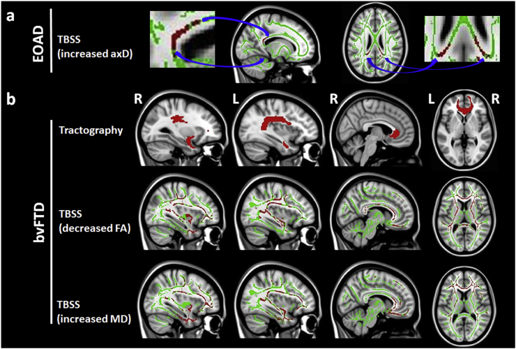
This prospective study explored whether an approach combining structural [cortical thickness and white matter (WM) microstructure] and resting state functional MRI can aid differentiation between 62 early onset Alzheimer's disease (EOAD) and 27 behavioural variant of frontotemporal dementia (bvFTD) patients. Random forest and receiver operator characteristic curve analyses assessed the ability of MRI in classifying the two clinical syndromes. All patients showed a distributed pattern of brain alterations relative to controls. Compared to bvFTD, EOAD patients showed bilateral inferior parietal cortical thinning and decreased default mode network functional connectivity. Compared to EOAD, bvFTD patients showed bilateral orbitofrontal and temporal cortical thinning, and WM damage of the corpus callosum, bilateral uncinate fasciculus, and left superior longitudinal fasciculus. Random forest analysis revealed that left inferior parietal cortical thickness (accuracy 0.78, specificity 0.76, sensitivity 0.83) and WM integrity of the right uncinate fasciculus (accuracy 0.81, specificity 0.96, sensitivity 0.43) were the best predictors of clinical diagnosis. The combination of cortical thickness and DT MRI measures was able to distinguish patients with EOAD and bvFTD with accuracy 0.82, specificity 0.76, and sensitivity 0.96. The diagnostic ability of MRI models was confirmed in a subsample of patients with biomarker-based clinical diagnosis. Multiparametric MRI is useful to identify brain alterations which are specific to EOAD and bvFTD. A severe cortical involvement is suggestive of EOAD, while a prominent WM damage is indicative of bvFTD.

Keywords: ACE-R, Addenbrooke's Cognitive Examination-revised; Behavioural variant of frontotemporal dementia; CC, corpus callosum; CSF, cerebrospinal fluid; Cortical thickness; DMN, default mode network; DT, diffusion tensor; Diagnosis; EOAD, early onset Alzheimer's disease; Early onset Alzheimer's disease; GM, grey matter; IC, independent component; ILF, inferior longitudinal fasciculus; LOAD, late onset Alzheimer's disease; MNI, Montreal Neurological Institute; NVI, Normalized Variable Importance; RS fMRI, resting state functional MRI; RSN, resting state network; Resting state functional MRI; SLF, superior longitudinal fasciculus; TFCE, threshold-free cluster enhancement; WM, white matter; White matter (WM) damage; bvFTD, behavioural variant frontotemporal dementia.


Figures

Fig. 1
Fig. 1
Cortical thickness findings. Patterns of regional cortical thinning of early onset Alzheimer's disease (EOAD) patients compared to cases with the behavioural variant of frontotemporal dementia (bvFTD) (blue) and vice versa (green). Results are overlaid on the Montreal Neurological Institute standard brain and shown at p < 0.05 False Discovery Rate-corrected for multiple comparisons. R = right; L = left.
Fig. 2
Fig. 2
Diffusion tensor MRI findings. A) Tract-Based Spatial Statistics (TBSS) results in early onset Alzheimer's disease (EOAD) patients compared to cases with the behavioural variant of frontotemporal dementia (bvFTD). The significant voxels are magnified for a better view. B) Tractography and TBSS findings in bvFTD patients relative to EOAD cases. TBSS results are shown in red (fractional anisotropy [FA], mean diffusivity [MD] and axial diffusivity [axD]) on the white matter (WM) skeleton (light green) and displayed on the sagittal and axial sections of the Montreal Neurological Institute (MNI) standard brain in neurological convention (p < 0.05 corrected for multiple comparisons at the cluster level using the threshold-free cluster enhancement option). WM tracts showing altered diffusion tensor MRI values are shown in red on the MNI standard brain in neurological convention (p < 0.05, false discovery rate-corrected for multiple comparisons). R = right; L = left.
Fig. 3
Fig. 3
Random forest analysis. Normalized variable importance of the five best MRI variables in distinguishing patients with early onset Alzheimer's disease (EOAD) and the behavioural variant of frontotemporal dementia (bvFTD), among A) cortical thickness measures (blue: greater cortical thinning in EOAD relative to bvFTD; green: greater cortical thinning in bvFTD relative to EOAD) and B) tractography measures (red; all regions showed greater damage in bvFTD compared to EOAD).
Fig. A.1 legend
Fig. A.1 legend
. Diffusion tensor MRI findings. Tract-Based Spatial Statistics (TBSS) results in (A) early onset Alzheimer's disease (EOAD) patients compared to healthy controls; and (B) behavioural frontotemporal dementia (bvFTD) patients compared to healthy controls. Fractional anisotropy is shown in red and mean diffusivity in blue on the white matter skeleton (light green) and displayed on the sagittal and axial sections of the Montreal Neurological Institute (MNI) standard brain in neurological convention (p < 0.05 corrected for multiple comparisons at the cluster level using the threshold-free cluster enhancement option). R = right; L = left.
Fig. A.2 legend
Fig. A.2 legend
. Resting state functional connectivity findings. A) Spatial map of the default mode network (DMN) is shown in cyan on the Montreal Neurological Institute (MNI) standard brain in neurological convention. B) Regions of decreased DMN functional connectivity in early onset Alzheimer's disease (EOAD) patients compared to cases with the behavioural variant of frontotemporal dementia (bvFTD). Results are overlaid on the MNI standard brain in neurological convention and shown at p < 0.05, family-wise error corrected for multiple comparisons (FWE). Colour bar denotes p values. R = right; L = left.

References

    1. Agosta F., Galantucci S., Magnani G., Marcone A., Martinelli D., Antonietta Volonte M., Riva N., Iannaccone S., Ferraro P.M., Caso F., Chio A., Comi G., Falini A., Filippi M. MRI signatures of the frontotemporal lobar degeneration continuum. Hum. Brain Mapp. 2015;36:2602–2614. - PMC - PubMed
    1. Alladi S., Xuereb J., Bak T., Nestor P., Knibb J., Patterson K., Hodges J.R. Focal cortical presentations of Alzheimer's disease. Brain. 2007;130:2636–2645. - PubMed
    1. Andersson J.L., Jenkinson M., Smith S. Technical Report. FMRIB Centre; Oxford, United Kingdom: 2007. Non-linear registration, aka spatial normalisation.
    1. Beckmann C.F., DeLuca M., Devlin J.T., Smith S.M. Investigations into resting-state connectivity using independent component analysis. Philos. Trans. R. Soc. Lond. Ser. B Biol. Sci. 2005;360:1001–1013. - PMC - PubMed
    1. Behrens T.E., Berg H.J., Jbabdi S., Rushworth M.F., Woolrich M.W. Probabilistic diffusion tractography with multiple fibre orientations: what can we gain? NeuroImage. 2007;34:144–155. - PMC - PubMed

Publication types

MeSH terms