

Accuracy of quantitative echocardiographic measures of right ventricular function as compared to cardiovascular magnetic resonance
- PMID: 28616541
- PMCID: PMC5454157
- DOI: 10.1016/j.ijcha.2016.05.007
Accuracy of quantitative echocardiographic measures of right ventricular function as compared to cardiovascular magnetic resonance
Abstract
Background: Many echocardiographic parameters have been proposed to evaluate right ventricular (RV) systolic function. We comprehensively assessed a wide range of quantitative echocardiographic parameters in a single cohort compared with same-day cardiovascular magnetic resonance (CMR).
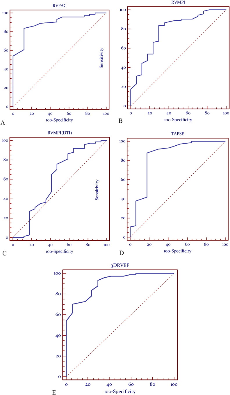
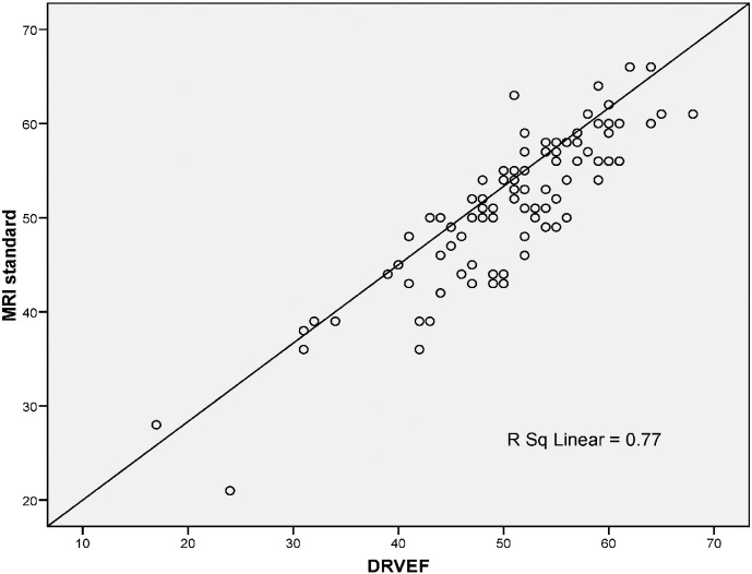
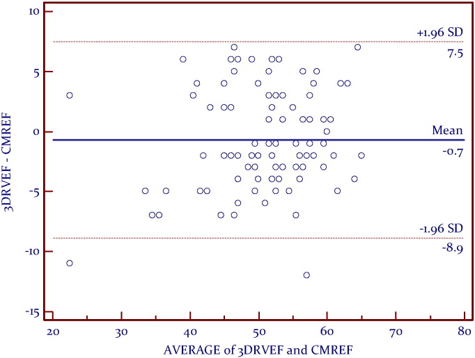
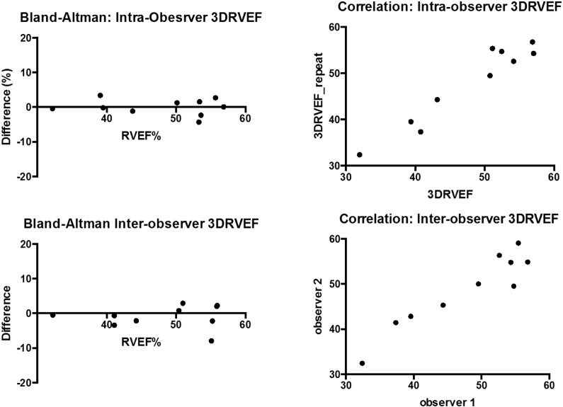
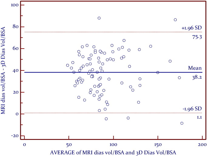
Methods and results: 92 subjects were examined prospectively: Group 1 consisted of 46 healthy controls (21 males, 33.4 ± 11.4 years), Group 2 consisted of 46 patients (20 males, 38.5 ± 18.9 years) undergoing RV functional assessment by CMR (1.5 T). Echocardiography was performed on the same day as CMR; fractional area change (RVFAC), myocardial performance index via spectral Doppler (RVMPI), RVMPI via Doppler tissue imaging (RVMPI-DTI), peak systolic myocardial velocity by DTI (RVSm), tricuspid annular plane systolic excursion (TAPSE), speckle tracking strain, and three dimensional right ventricular ejection fraction (3DE-RV). Linear regression, Bland-Altman and receiver-operator-characteristic (ROC) analyses were performed. At ROC analysis, the most predictive echocardiographic methods were; RVFAC (AUC = 0.892), RVMPI (AUC 0.785), TAPSE (AUC 0.849) and 3DE-RV (AUC 0.909). 3DE-RV appeared the most accurate compared to CMR, although underestimated true RV volumes.
Conclusion: As compared to CMR; 3DE-RV, RVFAC, TAPSE and RVMPI were the most reliable predictors of RV function. These parameters can be recommended for clinical use.
Keywords: 3DE, three dimensional echocardiography; 3DE-RV, three-dimensional echo right ventricular ejection fraction; CMR, cardiovascular magnetic resonance; DTI, Doppler tissue imaging; EF, ejection fraction; Echocardiography; IVCT, isovolumic contraction time; IVRT, isovolumic relaxation time; LV, left ventricle; MPI, myocardial performance index; Magnetic resonance imaging; RV, right ventricular; RVOT, right ventricular outflow tract; RVSm, peak systolic myocardial velocity; RVSm, s prime: right ventricular peak systolic myocardial velocity; Right ventricle; Right ventricular function; SR, strain rate; TAPSE, tricuspid annular peak systolic excursion; TOF, tetralogy of Fallot; TR, tricuspid regurgitation; ε, strain.
Figures
References
-
- D'Andrea A. Right ventricular myocardial dysfunction in adult patients late after repair of tetralogy of fallot. Int. J. Cardiol. 2004;94(2–3):213–220. - PubMed
-
- Haddad F. Right ventricular myocardial performance index predicts perioperative mortality or circulatory failure in high-risk valvular surgery. J. Am. Soc. Echocardiogr. 2007;20(9):1065–1072. - PubMed
-
- Kjaergaard J., Sogaard P., Hassager C. Quantitative echocardiographic analysis of the right ventricle in healthy individuals. J. Am. Soc. Echocardiogr. 2006;19(11):1365–1372. - PubMed
-
- Morner S. Right ventricular dysfunction in hypertrophic cardiomyopathy as evidenced by the myocardial performance index. Int. J. Cardiol. 2008;124(1):57–63. - PubMed
-
- Schwerzmann M. Comparison of echocardiographic and cardiac magnetic resonance imaging for assessing right ventricular function in adults with repaired tetralogy of fallot. Am. J. Cardiol. 2007;99(11):1593–1597. - PubMed
LinkOut - more resources
Full Text Sources
Other Literature Sources
Research Materials
