Case Reports
. 2017 May 16:2:49.
doi: 10.21037/tgh.2017.04.11. eCollection 2017.

Splenic laceration after routine colonoscopy, a case report of a rare iatrogenic complication

Affiliations
Case Reports

Splenic laceration after routine colonoscopy, a case report of a rare iatrogenic complication

Shuo Li et al. Transl Gastroenterol Hepatol. .

Abstract

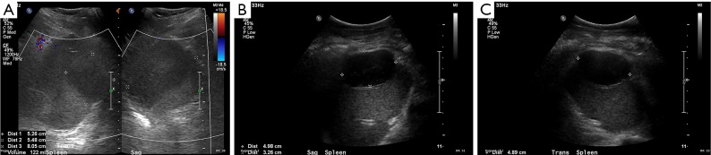
Colonoscopy is a common and routine procedure performed in the United States, most commonly performed for screening of colorectal cancer. Although colonoscopy is considered a safe procedure, it is associated with complications including intestinal hemorrhage and perforation. Splenic trauma, such as laceration or even complete rupture is a rarely reported, but potentially fatal complication if undetected. We present a case of splenic laceration with subcapsular hematoma status post routine colonoscopy. Fortunately, patient was able to be managed medically, without further operative intervention. We will also review the available literature related to this rare iatrogenic complication.

Keywords: Colonoscopy; radiology; splenic laceration.


Conflict of interest statement

Conflicts of Interest: The authors have no conflicts of interest to declare.

Figures

Figure 1
Figure 1
Colonoscopic images showing sigmoid diverticula (A) and a 5 mm sessile polyp at 30 cm (B) from the anal verge.
Figure 2
Figure 2
Contrast-enhanced CT performed in the ED two weeks after the colonoscopy. Axial as well as coronal and sagittal reformatted images showing a grade II splenic injury. A 1.8 cm, linear hypodense line is seen at the lateral aspect of the spleen on the coronal reformatted image (arrow). There is no evidence of extracapsular hemorrhage, such as fluid in the left paracolic gutter.
Figure 3
Figure 3
Follow up imaging of the spleen 7 days after patient discharged from the ED using color Doppler ultrasound (A) demonstrating a hypoechoic subcapsular splenic hematoma (marked by calipers) without evidence of active extravasation. Further follow up imaging of the spleen 14 days after patient discharge from the ED using grayscale ultrasound showing continued retraction of the subcapsular hematoma. Again, there is no evidence of extravasation into the hematoma on color Doppler (not shown).

References

    1. 1Seeff LC, Richards TB, Shapiro JA, et al. How many endoscopies are performed for colorectal cancer screening? results from CDC’s survey of endoscopic capacity. Gastroenterology 2004;127:1670-77. 10.1053/j.gastro.2004.09.051 - DOI - PubMed
    1. Centers for Disease Control and Prevention (CDC) Vital signs: Colorectal cancer screening test use--united states, 2012. MMWR Morb Mortal Wkly Rep 2013;62:881-8. - PMC - PubMed
    1. Whitlock EP, Lin JS, Liles E, et al. Screening for colorectal cancer: A targeted, updated systematic review for the US preventive services task force. Ann Intern Med 2008;149:638-58. 10.7326/0003-4819-149-9-200811040-00245 - DOI - PubMed
    1. Fisher DA, Maple JT, Ben-Menachem T, et al. Complications of colonoscopy. Gastrointest Endosc 2011;74:745-52. 10.1016/j.gie.2011.07.025 - DOI - PubMed
    1. Rabeneck L, Paszat LF, Hilsden RJ, et al. Bleeding and perforation after outpatient colonoscopy and their risk factors in usual clinical practice. Gastroenterology 2008;135:1899-906. 10.1053/j.gastro.2008.08.058 - DOI - PubMed

Publication types

LinkOut - more resources