Review
. 2017 Aug;8(4):377-392.
doi: 10.1007/s13244-017-0558-1. Epub 2017 Jun 14.

Liver segmentation: indications, techniques and future directions

Affiliations
Review

Liver segmentation: indications, techniques and future directions

Akshat Gotra et al. Insights Imaging. 2017 Aug.

Abstract

Objectives: Liver volumetry has emerged as an important tool in clinical practice. Liver volume is assessed primarily via organ segmentation of computed tomography (CT) and magnetic resonance imaging (MRI) images. The goal of this paper is to provide an accessible overview of liver segmentation targeted at radiologists and other healthcare professionals.

Methods: Using images from CT and MRI, this paper reviews the indications for liver segmentation, technical approaches used in segmentation software and the developing roles of liver segmentation in clinical practice.

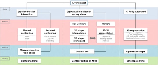
Results: Liver segmentation for volumetric assessment is indicated prior to major hepatectomy, portal vein embolisation, associating liver partition and portal vein ligation for staged hepatectomy (ALPPS) and transplant. Segmentation software can be categorised according to amount of user input involved: manual, semi-automated and fully automated. Manual segmentation is considered the "gold standard" in clinical practice and research, but is tedious and time-consuming. Increasingly automated segmentation approaches are more robust, but may suffer from certain segmentation pitfalls. Emerging applications of segmentation include surgical planning and integration with MRI-based biomarkers.

Conclusions: Liver segmentation has multiple clinical applications and is expanding in scope. Clinicians can employ semi-automated or fully automated segmentation options to more efficiently integrate volumetry into clinical practice.

Teaching points: • Liver volume is assessed via organ segmentation on CT and MRI examinations. • Liver segmentation is used for volume assessment prior to major hepatic procedures. • Segmentation approaches may be categorised according to the amount of user input involved. • Emerging applications include surgical planning and integration with MRI-based biomarkers.

Keywords: Automated; Computed tomography; Liver; Magnetic resonance imaging; Segmentation; Volumetry.


Figures

Fig. 1
Fig. 1
Variability of liver shape and size. Livers of different shape and volume may have similar cranial-caudal length, as demonstrated with these examples of three different patients. This observation highlights the limitation of reporting a one-dimensional measure of length, a well-entrenched practice, as a surrogate measure of liver volume
Fig. 2
Fig. 2
Types of major hepatectomy. White segments are planned for surgical resection. a Complete right hepatectomy. b Extended right hepatectomy. c Complete left hepatectomy. d Extended left hepatectomy. Figure adapted from the Brisbane 2000 Terminology of Liver Anatomy and Resections [12, 13]
Fig. 3
Fig. 3
Schematic of functional liver remnant (FLR) over total liver volume (TLV) ratio prior to hepatectomy. To be considered safely resectable prior to hepatectomy, the FLR/TLV ratio must be >20% in underlying normal livers, >30% in moderately diseased livers and >40% in cirrhotic livers
Fig. 4
Fig. 4
Future liver remnant volume calculation in normal liver prior to right hepatectomy. a Axial enhanced CT image showing colorectal liver metastasis involving right posterior segments (VI and VII). b Resection diagram shows the intended complete right hepatectomy surgery planned. c Three-dimensional rendered image showing surgical planning for complete right hepatectomy. FLR/TLV ratio was estimated to be 33%. d Axial unenhanced CT image of the same patient shortly after complete right hepatectomy. Actual FLR/TLV ratio was calculated to be 36%. Figure courtesy of Dr. Vandenbroucke-Menu; created with 3DVSP (IRCAD, Strasbourg, France)
Fig. 5
Fig. 5
Portal vein embolisation prior to right hepatectomy. a Axial enhanced CT image shows colorectal liver metastasis involving segments V, VI, VII (only VII shown). b Final portogram of embolised portal vein branches in segments V through VIII using a Lipiodol-glue mixture. c Axial enhanced CT image obtained 1 month after right PVE showing hypertrophy of future liver remnant. d Axial enhanced CT image of the same patient after right hepatectomy
Fig. 6
Fig. 6
Size incompatibility after living donor liver transplantation: both the donor and the recipient suffered transient hepatic insufficiency. a Axial enhanced CT image of a 26-year-old living liver donor. The total liver volume (TLV) was 1,754 mL. The donated liver volume was 980 mL and the residual liver volume was 774 mL (44.2% of the TLV). b Diagram showing the intended right split liver surgery planned for living donor liver transplantation. c Post-liver transplantation axial enhanced-CT image showing hypertrophied left liver of the donor. d Post-liver transplantation axial enhanced-CT image of a 53-year-old man who was the recipient of the right liver transplant. Although pre-transplant volumetry calculations seemed to indicate that the liver size was appropriate, the patient still developed small-for-size syndrome requiring ligature of the splenic artery. The cause was likely multifactorial. The donor developed transient biological hepatic insufficiency that resolved with supportive management
Fig. 7
Fig. 7
Workflows of various segmentation strategies. The schematic breaks down liver segmentation methods into truly manual, contour optimisation, semi-automated, and fully automated workflows. Most workflows require a combination of two-dimensional or three-dimensional initialisation, refinement and editing techniques. VOI volume of interest, MPR multi-planar reconstruction
Fig. 8
Fig. 8
Manual segmentation of the liver. a Manual segmentation of the liver performed by contouring of pixels of the liver boundary on CT image. Image obtained using Osirix image post-processing software (Osirix Foundation, Geneva, Switzerland). b Volume of the liver is obtained based on pixel size and slice spacing [13]
Fig. 9
Fig. 9
Active contours technique. a Image analyst roughly contours the liver using a cursor. b Contour evolves based on salient image features. c The contour snaps to the true liver contour [13]
Fig. 10
Fig. 10
Livewire technique. a User sets the “seed point” by clicking on the liver boundary. b As the cursor is moved, the boundary behaves like a livewire, connecting the seed point to the cursor. c The free point is placed along the liver boundary, and a minimal cost path is generated [13]
Fig. 11
Fig. 11
Seeded region-growing technique. a Seeds are positioned inside the regions of interest by user. b Pixels are iteratively aggregated if their intensity is similar to those already tagged. c The liver parenchyma is segmented
Fig. 12
Fig. 12
Statistical shape models. To restrict the segmentation to a set of admissible liver shapes, a shape database is compiled, from which any new liver shape is expressed by a set of parameters called modes of variation. The various modes of variation (roughly 30 modes) are adjusted to fit the liver shape on image features. Statistical shape models impose hard restriction on the segmentation outcome by integrating prior shape. However, training data cannot capture all variations and therefore are sometimes too limiting to accurately model specific livers [13]
Fig. 13
Fig. 13
Imaging pitfalls which may degrade liver segmentation on MRI. Axial T1-weighted fat-saturated images with contrast injection depict the following artefacts: a Severe motion artefact. b Partial volume averaging of the liver parenchyma with the gallbladder (arrows). c Ghost artefact with the aorta (arrow). d Inhomogeneous fat saturation (white arrows) and fat-water swap in the liver (arrowheads) [13]
Fig. 14
Fig. 14
Imaging pitfalls which may limit liver segmentation on CT. a Axial enhanced CT image of a 62-year-old woman shows indistinct liver-spleen boundaries (arrows). b Axial enhanced CT image of a 47-year-old man depicts segmentation challenges caused by ill-defined and non-continuous borders found near the liver dome (arrows). c Axial enhanced CT image of a 73-year-old man shows partial volume averaging between the left liver and the heart (arrows) [13]
Fig. 15
Fig. 15
Liver subsegmentation according to vascular anatomy. Axial enhanced CT showing the segmented a hepatic arterial, b portal venous and c hepatic venous structures. Three-dimensional rendering of the same liver showing the corresponding segmented d arterial, e portal venous and f hepatic venous structures [13]
Fig. 16
Fig. 16
Virtual surgical planning. a Axial enhanced CT image shows a right liver metastasis centred in segment V (arrow). The patient also had a metastasis involving segment VII (not shown). b Axial enhanced CT image of a different patient shows a left liver metastasis in segment III (arrow). c Three-dimensional rendering image shows surgical planning for complete right hepatectomy including tumour and hepatic structures in patient from a. d Three-dimensional rendered image shows surgical planning for segmentectomy of segment III for patient in b. Residual hepatic liver volume after both procedures was estimated to be 27%. Right portal embolisation was thus performed before right hepatectomy. Figure courtesy of Dr. Vandenbroucke-Menu; created with 3DVSP (IRCAD, Strasbourg, France)

References

    1. Castell DO, O’Brien KD, Muench H, Chalmers TC. Estimation of liver size by percussion in normal individuals. Ann Intern Med. 1969;70(6):1183–1189. doi: 10.7326/0003-4819-70-6-1183. - DOI - PubMed
    1. Vauthey JN, Chaoui A, Do KA, et al. Standardized measurement of the future liver remnant prior to extended liver resection: methodology and clinical associations. Surgery. 2000;127(5):512–519. doi: 10.1067/msy.2000.105294. - DOI - PubMed
    1. Martel G, Cieslak KP, Huang R, et al. Comparison of techniques for volumetric analysis of the future liver remnant: implications for major hepatic resections. HPB (Oxford) 2015;17(12):1051–1057. doi: 10.1111/hpb.12480. - DOI - PMC - PubMed
    1. Heymsfield SB, Fulenwider T, Nordlinger B, Barlow R, Sones P, Kutner M. Accurate measurement of liver, kidney, and spleen volume and mass by computerized axial tomography. Ann Intern Med. 1979;90(2):185–187. doi: 10.7326/0003-4819-90-2-185. - DOI - PubMed
    1. Nakayama Y, Li Q, Katsuragawa S, et al. Automated hepatic volumetry for living related liver transplantation at multisection CT. Radiology. 2006;240(3):743–748. doi: 10.1148/radiol.2403050850. - DOI - PubMed