. 2017 May;9(2):206-214.
doi: 10.1111/os.12331. Epub 2017 Jun 14.

Treatment for Thoracic Ossification of Posterior Longitudinal Ligament with Posterior Circumferential Decompression

Affiliations

Treatment for Thoracic Ossification of Posterior Longitudinal Ligament with Posterior Circumferential Decompression

Zhao-Wan Xu et al. Orthop Surg. 2017 May.

Abstract

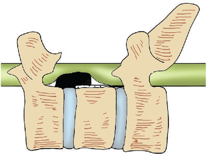
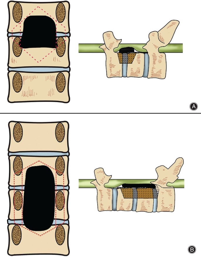
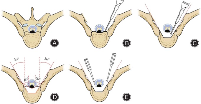
Objective: To report the results of the posterior approach for thoracic ossification of posterior longitudinal ligament (TOPLL) by using a special "L" osteotome.

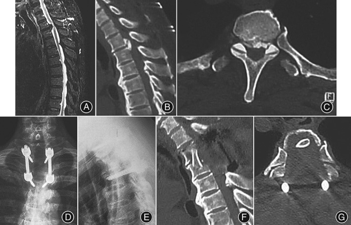
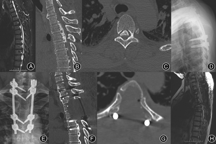
Methods: The present study enrolled 16 consecutive patients (9 men and 7 women) between May 2009 and September 2013. All patients underwent a posterior circumferential decompression osteotomy and segmental instrumentation with interbody fusion. The mean age at surgery was 57.3 years (range, 37-68 years). Patients' data, clinical manifestation, blood loss, length of surgery, complications, visual analog scale (VAS), Japanese Orthopedic Association (JOA), and Frankel grading system before and after surgery were collected and evaluated, retrospectively.

Results: The average follow-up period was 30 ± 19 months (range, 12-50 months). All patients were successfully treated with posterior compression and segmental instrumentation with interbody fusion. The average operation time was 261.6 ± 51.3 min (range, 190-310 min). The mean blood loss was 980.3 ± 370.5 mL (range, 600-2100 mL). All patients had subjective improvement of motor power and gait. Average preoperative and postoperative JOA scores were 4.2 ± 1.7 and 7.8 ± 2.5 points, respectively. Differences in the overall JOA scores showed significant postoperative improvement. At the last follow-up, all patients improved either by one or two Frankel grades. There was a significant difference between preoperative VAS scores and those 3 months after surgery (P < 0.05). No significant difference was observed between the 3-month and 12-month results (P > 0.05). Cerebrospinal fluid (CSF) leakage occurred in 3 patients. Acute neurological deterioration was encountered postoperatively in 1 patient.

Conclusion: Treatment with posterior transpedicular osteotomy and circumferential decompression was found to be safe, effective, reliable, and technically feasible, and keeping the thoracic cavity intact avoids many shortcomings of anterior surgery and results in a satisfactory spinal decompression.

Keywords: Circumferential decompression; Internal fixation; Ossification of the posterior longitudinal ligament; Osteotomy.


Figures

Figure 1
Figure 1
Diagram of the “triangular safe area,” where normal and ossifying posterior longitudinal ligament is cut off. As the dural sac was compressed by the ossification of the posterior longitudinal ligament (OPLL), there was a potential triangular space at the OPLL ends, the posterior edge of the vertebral body, and the compressed dural sac, known as the “triangular safe area.”
Figure 2
Figure 2
Osteotomy scope in coronal and sagittal views of thoracic ossification of posterior longitudinal ligament (TOPLL). (A) Head end of OPLL is in middle–lower part of pedicle and the “triangular safe area” is not completely blocked; the pedicle can be retained in osteotomy. Invasion of the tail end of the OPLL to the transverse diameter of the spinal canal is 100%, and involves the bilateral pedicle inner wall. The osteotomy requires removing the corresponding pedicle. The exterior and inner walls of the pedicle are the exterior and inner boundaries of the osteotomy. (B) Head end of OPLL is in the middle–upper part of the pedicle, and the “triangular safe area” is blocked by the pedicle. Thus, the procedure requires partial or total pedicle removal to expose the “triangular safe area”. Resection of pedicle in the middle area of OPLL is necessary. The tail end of the OPLL is in the middle–upper part of the pedicle. The “triangular safe area” is not completely blocked, and the pedicle can be retained as part of the osteotomy procedure.
Figure 3
Figure 3
Schematic diagram of thoracic transpedicle osteotomy and circumferential decompression. (A) Diagram of thoracic ossification of posterior longitudinal ligament (TOPLL); (B) both lamina and bilateral articular processes are removed and the bilateral pedicles are abraded to the posterior edge of the vertebral body. The costotransverse articulations are retained, while the dural sac, nerve root, and “triangular safe area” are exposed; (C) transpedicle osteotomy is performed, where the shadow shows the resected part; (D) a “culvert” is generated after resection of one‐quarter to half of the posterior vertebral body. The OPLL is floating above the culvert; and (E) the OPLL is cut off using an “L” model osteotome and pushed into the “culvert”.
Figure 4
Figure 4
Imaging of the preoperative and postoperative surgery levels (female, 57 years old). (A) Preoperative MRI shows a unisegmental bird‐beak thoracic ossification of posterior longitudinal ligament (TOPLL), with significant compression at the T 2–3 spinal cord plane; (B) preoperative sagittal CT scan shows a unisegmental isolated T‐OPLL at T 2–3; and (C) preoperative axial CT scan shows the tail end of OPLL is at the T 3 pedicle. The invasion of the OPLL to the transverse diameter of the spinal canal is 100%, involving the inner wall of the bilateral pedicle; (D, E) postoperative lateral side X‐ray photograph shows pedicle screw internal fixation at T 2 and T 4, with a good fixation position; and (F) postoperative sagittal CT scan shows the “culvert” has been removed. (G) A postoperative axial CT scan shows total removal of the T 3 OPLL; complete decompression of spinal cord is achieved, and costotransverse articulations were retained.
Figure 5
Figure 5
Imaging of the preoperative and postoperative surgery levels (male, 46 years old). (A) Preoperative sagittal T 2‐weighted MRI shows a mixed T‐OPLL, with significant compression in spinal cord planes of T 2–3, T 3–4, and T 5–6; (B) preoperative sagittal CT scan shows continuous OPLL in planes of T 2–3, T 3–4, and T 5–6; (C) preoperative axial CT scan reveals that the OPLL tail end is at the T 5 pedicle level, the occupation ratio of the OPLL on the transverse diameter of spinal canal is less than 100%, and the bilateral pedicular inner wall is not involved; (D, E) postoperative lateral side X‐ray photography reveals pedicular screw internal fixation at T 1, T 2, T 4, and T 5 levels, with a good fixation position; (F) postoperative sagittal CT scan shows complete resection of the OPLL; (G) postoperative axial CT scan shows a total resection of OPLL at T 3, with a complete decompression of the spinal cord, while retaining the costovertebral joints; (H) a postoperative sagittal T 2‐weighted MRI shows the total resection of the OPLL and complete decompression of the spinal cord.

References

    1. Matsumoto M, Chiba K, Toyama Y, et al. Surgical results and related factors for ossification of posterior longitudinal ligament of the thoracic spine: a multi‐institutional retrospective study. Spine (Phila Pa 1976), 2008, 33: 1034–1041. - PubMed
    1. Yang C, Bi Z, Fu C, Zhang Z. A modified decompression surgery for thoracic myelopathy caused by ossification of posterior longitudinal ligament: a case report and literature review. Spine (Phila Pa 1976), 2010, 35: E609–E613. - PubMed
    1. Kojima T, Waga S, Kubo Y, Matsubara T. Surgical treatment of ossification of the posterior longitudinal ligament in the thoracic spine. Neurosurgery, 1994, 34: 854–858. - PubMed
    1. Hanai K, Ogikubo O, Miyashita T. Anterior decompression for myelopathy resulting from thoracic ossification of the posterior longitudinal ligament. Spine (Phila Pa 1976), 2002, 27: 1070–1076. - PubMed
    1. Tsuzuki N, Hirabayashi S, Abe R, Saiki K. Staged spinal cord decompression through posterior approach for thoracic myelopathy caused by ossification of posterior longitudinal ligament. Spine (Phila Pa 1976), 2001, 26: 1623–1630. - PubMed

LinkOut - more resources