. 2017 Jun 15;6(2):43.
doi: 10.3390/antiox6020043.

NOX2-Induced Activation of Arginase and Diabetes-Induced Retinal Endothelial Cell Senescence

Affiliations

NOX2-Induced Activation of Arginase and Diabetes-Induced Retinal Endothelial Cell Senescence

Modesto Rojas et al. Antioxidants (Basel). .

Abstract

Increases in reactive oxygen species (ROS) and decreases in nitric oxide (NO) have been linked to vascular dysfunction during diabetic retinopathy (DR). Diabetes can reduce NO by increasing ROS and by increasing activity of arginase, which competes with nitric oxide synthase (NOS) for their commons substrate l-arginine. Increased ROS and decreased NO can cause premature endothelial cell (EC) senescence leading to defective vascular repair. We have previously demonstrated the involvement of NADPH oxidase 2 (NOX2)-derived ROS, decreased NO and overactive arginase in DR. Here, we investigated their impact on diabetes-induced EC senescence. Studies using diabetic mice and retinal ECs treated with high glucose or H₂O₂ showed that increases in ROS formation, elevated arginase expression and activity, and decreased NO formation led to premature EC senescence. NOX2 blockade or arginase inhibition prevented these effects. EC senescence was also increased by inhibition of NOS activity and this was prevented by treatment with a NO donor. These results indicate that diabetes/high glucose-induced activation of arginase and decreases in NO bioavailability accelerate EC senescence. NOX2-generated ROS contribute importantly to this process. Blockade of NOX2 or arginase represents a strategy to prevent diabetes-induced premature EC senescence by preserving NO bioavailability.

Keywords: NO; NOS; NOX2/NADPH oxidase 2; arginase 1; diabetic retinopathy; endothelial cell; oxidative stress; retina; senescence associated β-galactosidase.


Conflict of interest statement

The authors declare that there is no conflict of interest associated with this manuscript.

Figures

Figure 1
Figure 1
NADPH oxidase 2 (NOX2) deletion prevents diabetes-induced increases in arginase 1 expression and activity. Effects of NOX2 deletion on expression of arginase l (Arg1) protein (A) and arginase activity (B) in diabetic and control retinas. * p < 0.05 vs. control, # p < 0.05 vs. WT diabetic, Values are mean ± SEM, n = 4–5.
Figure 2
Figure 2
NOX2 deletion prevents diabetes-induced decreases in retinal nitric oxide (NO). Representative images and quantitation showing effects of NOX2 deletion on fluorescence for the NO indicator DAF-2DA (arrows) in diabetic and control retinas. Quantitative analysis showed significant reduction of NO levels in samples from WT diabetic retinas as compared to control retinas. NO was restored by deletion of NOX2. Pretreatment with the NOS inhibitor L-NAME significantly decreased NO in WT retinas. * p < 0.05 vs. WT control, # p < 0.05 vs. WT diabetic. Values are mean ± SEM, n = 4–5. Scale bar = 50 μm.
Figure 3
Figure 3
NOX2 deletion limits diabetes-induced increases in SA-β-gal activity. Representative images showing effects of diabetes on SA-β-gal activity in the inner retina. Retinas of WT diabetic mice showed numerous SA-β-gal positive cells (red arrows) in the inner retina. The SA-β-gal signal is largely absent in retinas from the NOX2−/− diabetic mice. Scale bar = 50 μm.
Figure 4
Figure 4
NOX2 blockade limits high glucose-induced increases in SA-β-gal activity. Representative images and statistical analysis showing effects of high glucose on SA-β-gal activity in retinal endothelial cells. Incubation of the cultures in high glucose (HG) media markedly increased the number of SA-β-gal-positive cells (arrows) as compared with the normal glucose (NG) controls. This was prevented by treatment with the NOX2-blocking peptide gp91ds-tat. Treatment with the scrambled control (SC) peptide did not alter the high glucose effect. * p < 0.0001 vs. NG, n = 6. Scale bar = 20 µm.
Figure 5
Figure 5
NOX2 blockade prevents high glucose-induced increases in oxidative stress. Representative images and statistical analysis of dihydroethidium (DHE) imaging showing effects of high glucose on reactive oxygen species (ROS) formation in endothelial cells. Treatment with high glucose (HG) markedly increased the DHE fluorescence as compared with the normal glucose controls (NG). Treatment with the NOX2-blocking peptide gp91ds-tat but not the scrambled control (SC) peptide blocked the HG-induced increase in ROS. * p < 0.05, n = 10. Scale bar = 20 µm.
Figure 6
Figure 6
NOX2 blockade prevents high glucose-induced increases in arginase 1 expression and arginase activity in retinal endothelial cells. Western blot and quantitation showing effects of the NOX2-blocking peptide gp91ds-tat on expression of arginase l (Arg1) protein (A) and arginase activity (B) in endothelial cells treated with high glucose (HG). HG substantially increased arginase 1 expression and arginase activity as compared to the normal glucose (NG) controls. These increases were blocked by bp91ds-tat but were not altered by the scrambled control (SC) peptide. * p < 0.05, n = 3–4.
Figure 7
Figure 7
NOX2 blockade prevents high glucose-induced decreases in endothelial cell nitric oxide. Representative images (A) and quantitation (B) showing effects of NOX2 blockade on fluorescence for the NO indicator DAF-2DA in endothelial cells maintained in high glucose (HG) or normal glucose (NG) media. Quantitative analysis showed significant reduction of NO levels in samples from HG-treated cells as compared to NG controls. NO was preserved in HG cultures treated with the NOX2-blocking peptide gp91ds-tat but not in cultures treated with the scrambled control (SC) peptide. Treatment with the NOS inhibitor L-NAME significantly decreased NO in the NG cultures but did not alter cell density as shown by phase contrast image. * p < 0.0001 vs. NG, # p < 0.05 vs. HG, n = 6. Analysis of NO accumulation in the conditioned media (C) confirmed a decrease in NO release in the HG cultures that was blocked by gp91ds-tat, but not by the scrambled control (SC). * p < 0.05 vs. NG, # p < 0.05 vs. HG, n = 4–6. Scale bar = 20 µm.
Figure 8
Figure 8
Hydrogen peroxide induces an increase in arginase activity. Treatment of retinal endothelial cells with hydrogen peroxide induced a dose-dependent increase in arginase activity as compared to control media. * p < 0.05 vs. control, n = 3–4.
Figure 9
Figure 9
Hydrogen peroxide induces an increase in SA-β-gal activity. Treatment of retinal endothelial cells with hydrogen peroxide or the NOS inhibitor l-NG-nitroarginine methyl ester (l-NAME) induced a significant increase in SA-β-gal activity (arrows). Treatment with the arginase inhibitor ABH blocked the hydrogen peroxide effect and treatment with the NO donor SNAP blocked the L-NAME effect. * p < 0.05 vs. control, n = 17. Scale bar = 30 µm.
Figure 10
Figure 10
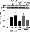
Hydrogen peroxide induces an increase in the cyclin-dependent kinase inhibitor p16INK4a. Western blot and quantitation showing effects of treatment with hydrogen peroxide or l-NAME in increasing expression of p16INK4a protein. These effects were blocked by the arginase inhibitor ABH (100 µM) or the NO donor SNAP (10 µM), * p < 0.05 vs. control, n = 3–4.
Figure 11
Figure 11
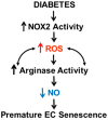
Hypothesis: Diabetes-induced activation of NOX2/NADPH oxidase promotes the development of premature endothelial cell senescence by a mechanism involving ROS-induced activation of arginase which amplifies ROS and decreases bioavailable NO.

References

    1. Zhang X., Saaddine J.B., Chou C.F., Cotch M.F., Cheng Y.J., Geiss L.S., Gregg E.W., Albright A.L., Klein B.E., Klein R. Prevalence of diabetic retinopathy in the United States, 2005–2008. JAMA. 2010;304:649–656. doi: 10.1001/jama.2010.1111. - DOI - PMC - PubMed
    1. Hammes H.P., Feng Y., Pfister F., Brownlee M. Diabetic retinopathy: Targeting vasoregression. Diabetes. 2011;60:9–16. doi: 10.2337/db10-0454. - DOI - PMC - PubMed
    1. Stitt A.W., Curtis T.M., Chen M., Medina R.J., McKay G.J., Jenkins A., Gardiner T.A., Lyons T.J., Hammes H.P., Simo R., et al. The progress in understanding and treatment of diabetic retinopathy. Prog. Retin. Eye Res. 2016;51:156–186. doi: 10.1016/j.preteyeres.2015.08.001. - DOI - PubMed
    1. Cohen S.R., Gardner T.W. Diabetic Retinopathy and Diabetic Macular Edema. Dev. Ophthalmol. 2016;55:137–146. - PMC - PubMed
    1. Goldberg M.F. Retinal detachment associated with proliferative retinopathies (sickle cell disease, retrolental fibroplasia and diabetes mellitus) Isr. J. Med. Sci. 1972;8:1447–1457. - PubMed

LinkOut - more resources