Review
. 2017 Jun 15;7(2):26.
doi: 10.3390/life7020026.

DNA Protection Protein, a Novel Mechanism of Radiation Tolerance: Lessons from Tardigrades

Affiliations
Review

DNA Protection Protein, a Novel Mechanism of Radiation Tolerance: Lessons from Tardigrades

Takuma Hashimoto et al. Life (Basel). .

Abstract

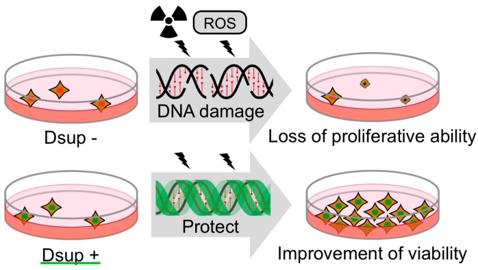
Genomic DNA stores all genetic information and is indispensable for maintenance of normal cellular activity and propagation. Radiation causes severe DNA lesions, including double-strand breaks, and leads to genome instability and even lethality. Regardless of the toxicity of radiation, some organisms exhibit extraordinary tolerance against radiation. These organisms are supposed to possess special mechanisms to mitigate radiation-induced DNA damages. Extensive study using radiotolerant bacteria suggested that effective protection of proteins and enhanced DNA repair system play important roles in tolerability against high-dose radiation. Recent studies using an extremotolerant animal, the tardigrade, provides new evidence that a tardigrade-unique DNA-associating protein, termed Dsup, suppresses the occurrence of DNA breaks by radiation in human-cultured cells. In this review, we provide a brief summary of the current knowledge on extremely radiotolerant animals, and present novel insights from the tardigrade research, which expand our understanding on molecular mechanism of exceptional radio-tolerability.

Keywords: damage suppressor (Dsup); extremophiles; radiotolerance; reactive oxygen species (ROS); tardigrade.


Conflict of interest statement

The authors declare no conflict of interest.

Figures

Figure 1
Figure 1
Scanning electron microscopy images of the extremotolerant tardigrade, R. varieornatus. Reproduced from [18].
Figure 2
Figure 2
Dsup reduced X-ray-induced DNA damage and improved viability of irradiated human cultured cells. (A) Quantitative comparison of DNA-break marker, γ-H2AX foci number among untransfected human cultured cells (Control), Dsup-expressing cells (Dsup), and Dsup-knockdown cells (Dsup + shDsup) under non-irradiated and 1 Gy X-ray irradiated conditions. ** p < 0.01, n.s. indicates not significant (Tukey–Kramer’s test). (B) Comparison of growth curves of untransfected cells (Control), Dsup-expressing cells (Dsup), and Dsup-knockdown cells (Dsup + shDsup) in irradiated conditions. Values represent mean ± s.d. Reproduced from [18].
Figure 3
Figure 3
The schematic structure of Dsup protein. This protein is composed of 445 amino acids (aa). The arrow indicates a predicted α-helix at the middle region and a bar in the C-terminus indicates a predicted nuclear localization signal.
Figure 4
Figure 4
Comparison of putative Dsup proteins in two tardigrade species. (A) Pairwise alignment of Dsup protein of R. varieornatus (Rv_Dsup; accession number = BAV59442) and putative Dsup protein of H. dujardini (Hd_Dsup, hypothetical protein BV898_01301, accession number = OQV24709). Identical residues and similar residues are shown in inverted boxes and shaded boxes, respectively. Predicted nuclear localization signals (NLS) are shown by red bars. Green shades indicate conserved alanine-rich regions detected by PROSITE protein pattern search. (B) Comparison of distribution profiles of hydrophobicity and charges between two tardigrade Dsup proteins. Hydrophobicity and charge distribution were analyzed using the ProtScale program at ExPASy and EMBOSS charge program, respectively.
Figure 4
Figure 4
Comparison of putative Dsup proteins in two tardigrade species. (A) Pairwise alignment of Dsup protein of R. varieornatus (Rv_Dsup; accession number = BAV59442) and putative Dsup protein of H. dujardini (Hd_Dsup, hypothetical protein BV898_01301, accession number = OQV24709). Identical residues and similar residues are shown in inverted boxes and shaded boxes, respectively. Predicted nuclear localization signals (NLS) are shown by red bars. Green shades indicate conserved alanine-rich regions detected by PROSITE protein pattern search. (B) Comparison of distribution profiles of hydrophobicity and charges between two tardigrade Dsup proteins. Hydrophobicity and charge distribution were analyzed using the ProtScale program at ExPASy and EMBOSS charge program, respectively.
Figure 5
Figure 5
Schematic model of DNA protection by Dsup protein from radiation damage. Radiation induces DNA breaks, which could interfere DNA replication and gene expression. Heavily damaged cells lose their proliferative ability and are destined for death. Dsup protein suppresses X-ray induced DNA damage depending on association with nuclear DNA, possibly through physical shielding from, or detoxification of, reactive oxygen species (ROS) generated through indirect radiation effects. Thereby, Dsup protein can improve the radiotolerance of cultured animal cells.

References

    1. Rothschild L.J., Mancinelli R.C. Life in extreme environments. Nature. 2001;409:1092–1101. doi: 10.1038/35059215. - DOI - PubMed
    1. Mayer G., Kauschke S., Rüdiger J., Stevenson P.A. Neural markers reveal a one-segmented head in tardigrades (water bears) PLoS ONE. 2013;8:e59090. doi: 10.1371/journal.pone.0059090. - DOI - PMC - PubMed
    1. Rebecchi L., Altiero T., Guidetti R. Anhydrobiosis: The extreme limit of desiccation tolerance. Invertebr. Surviv. J. 2007;4:65–81.
    1. Actual checklist of Tardigrada Species. [(accessed on 29 March 2017)];2015 Available online: http://www.tardigrada.modena.unimo.it/miscellanea/ActualchecklistofTardi....
    1. Vicente F., Bertolani R. Considerations on the taxonomy of the Phylum Tardigrada. Zootaxa. 2013;3626:245–248. doi: 10.11646/zootaxa.3626.2.2. - DOI - PubMed