. 2017 Aug 18;12(8):2085-2096.
doi: 10.1021/acschembio.7b00305. Epub 2017 Jun 28.

Antibodies Against Specific MUC16 Glycosylation Sites Inhibit Ovarian Cancer Growth

Affiliations

Antibodies Against Specific MUC16 Glycosylation Sites Inhibit Ovarian Cancer Growth

Thapi Dharma Rao et al. ACS Chem Biol. .

Abstract

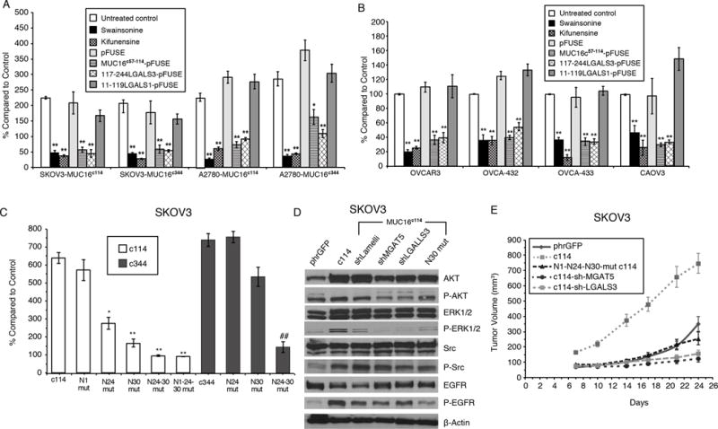
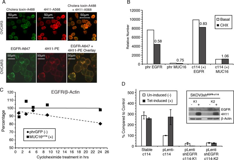
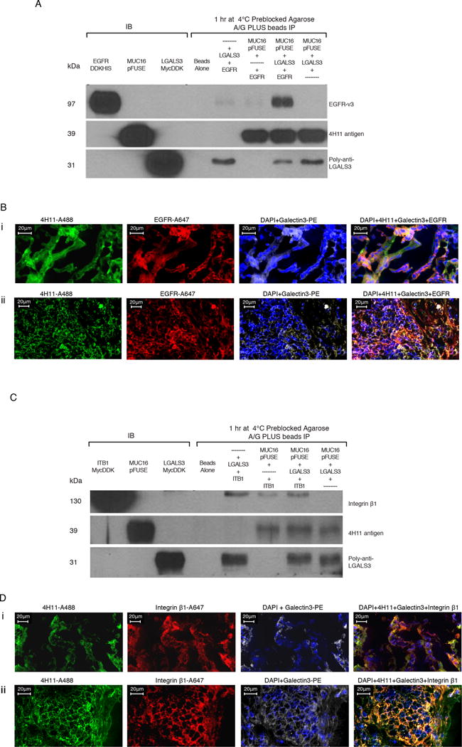
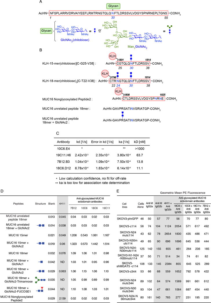
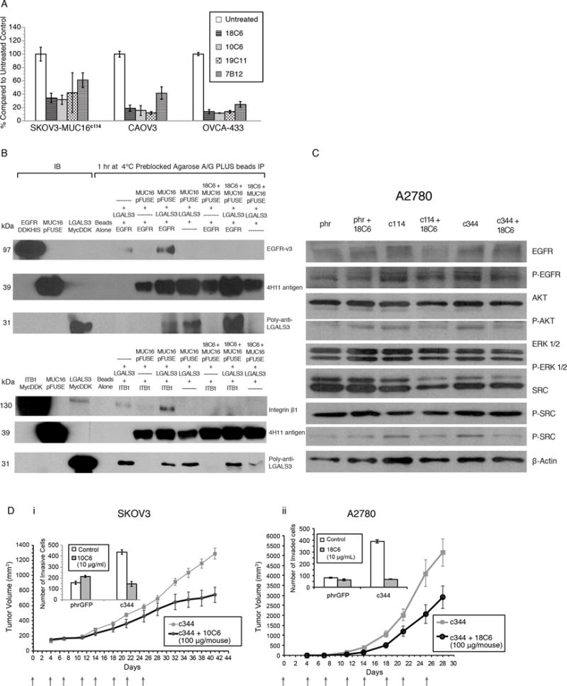
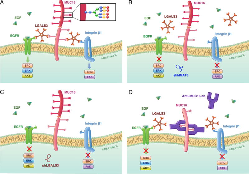
Expression of the retained C-terminal extracellular portion of the ovarian cancer glycoprotein MUC16 induces transformation and tumor growth. However, the mechanisms of MUC16 oncogenesis related to glycosylation are not clearly defined. We establish that MUC16 oncogenic effects are mediated through MGAT5-dependent N-glycosylation of two specific asparagine sites within its 58 amino acid ectodomain. Oncogenic signaling from the C-terminal portion of MUC16 requires the presence of Galectin-3 and growth factor receptors colocalized on lipid rafts. These effects are blocked upon loss of either Galectin-3 expression or activity MGAT5. Using synthetic MUC16 glycopeptides, we developed novel N-glycosylation site directed monoclonal antibodies that block Galectin-3-mediated MUC16 interactions with cell surface signaling molecules. These antibodies inhibit invasion of ovarian cancer cells, directly blocking the in vivo growth of MUC16-bearing ovarian cancer xenografts, elucidating new therapeutic modalities.


Conflict of interest statement

Conflict of Interest Statement

Memorial Sloan Kettering Cancer Center filed a patent application for MUC16 glycosylated ectodomain monoclonal antibodies on March 17, 2015, and the outcome is pending.

Figures

Figure 1
Figure 1. Effect of MUC16 Expression on SKOV3 and A2780 Ovarian Cancer Cells
A) MUC16 enhancement of matrigel invasion assay SKOV3 and A2780 cells depends on N-glycosylation. Cells were exposed to Swainsonine(1 ug/ml), Kifunensine (1ug/ml), control pFUSE protein, MUC16c57-114-pFUSE fusion protein, 117-244LGALS3-pFUSE, or 11-119LGALS1-pFUSE fusion protein (all 5 ug/ml) and compared to vector controls. Results (n=3) are expressed as percentages compared by paired t test to parent SKOV3 orA2780 phrGFP control after 48 h (Mean ± SE). (*=p,0.01; ** p< 0.0001) In both SKOV3 and A2780 cell lines, MUC16c114 or MUC16c344 cells were more invasive than the control phrGFP cells (** = p<0.0001), and these invasive properties were unaffected by exposure to the pFUSE vector-only protein. Each study was replicated ≥ 3 times. B) Matrigel invasion assay for wild-type ovarian cancer cell lines depends on Galectin-3. OVCAR3, OVCA-432, OVCA-433, and CAOV3 cells were exposed to Swainsosine (1 ug/ml), Kifunensin (1ug/m), control pFUSE protein, MUC16c57-114-pFUSE fusion protein, 117-244LGALS3-pFUSE, or 11-119LGALS1-pFUSE fusion protein (all 5 ug/ml). Invasion was measured in triplicate (n=3) and normalized against untreated control cells. Results (n=3) are expressed as percentages compared by paired t test to parental control cells after 48 h (Mean ± SE). (*=p,0.01; ** p< 0.0001) Two or more replicates were performed for each condition. C) Loss of proximal N-glycosylation sites impair matrigel invasion. SKOV3-MUC16c114 and SKOV3-MUC16c344 transfected cell lines were tested for MUC16-based increased invasion following N→A mutations of N-glycosylation sites at each of the N1, N24, and N30 positions. Invasion was measured in triplicate (n=3), with 3 or more independent replicates. Results are expressed as percentages compared to SKOV3phr cells without MUC16 expression. Results (n=3) are expressed as percentages compared by paired t test to parental control cells after 48 h (Mean ± SE). (*=p,0.01; ** p< 0.0001) D) MUC16-induced oncogene activation on AKT, MAPK, and SRC signaling pathways indicating MUC16 increased phosphorylation of AKT (S473), ERK1/2 (pT202/Y204), SRC(y416), and EGFR(pY1068) in SKOV3-MUC16c114 cells. MGAT5 (shMGAT5), Galectin-3 (shLGALS3) knockdowns, and N30A mutation all reduced MUC16c114-induced oncogene activation. E) MUC16 N-glycosylation–dependent tumor growth in vivo. In vivo growth of SKOV3-MUC16c114 expressing cell line was much more aggressive (p<0.0001 by paired t test) compared to the SKOV3-phrGFP control. However, SKOV3-MUC16(N1-N24-N30)mut-c114 glycosylation -impaired transfectants did not show any growth enhancement compared to SKOV3-phrGFP vector control tumors. SKOV-3-MUC16c114 cells with shRNA of LGALS3 or MGAT5 were also similar to control cells
Figure 2
Figure 2. MUC16 expression increases EGFR expression and stability
A) Cell surface MUC16 is localized in lipid rafts and co-localizes with EGFR. Cholera toxin localizes to lipid rafts on the cell surface (green label) (Alexa488) and co-localizes with the red-labeled anti-MUC16 (Alexa568) on the cell surface of OVCAR 3 cell line. In the same cell line, EGFR (Alexa647 green label) also co-localizes with MUC16 (Phycoerythrin). B) MUC16 increased EGFR expression. Cells were labeled wth anti-EGFR-A647 and relative number estimated by FACS geometric mean fluorescence. Relative cell surface EGFR expression was reduced to 58% of untreated levels upon 24 h of CHX exposure in SKOV3-phrGFP. In contrast, in the SKOV3-MUC16c114 cells, there was an increase in EGFR geometric mean fluorescence, which decreased to 83% of that of the control after CHX exposure. MUC16c114 mean fluorescence is not reduced by CHX. C) Expression of MUC16c114 stabilizes total EGFR after cycloheximide. SKOV3-cells with and without MUC16 expression were exposed to cycloheximide and expression of EGFR species were compared at various times. Densitometry of the EGFR/β-actin ratio illustrates that there is a steady loss of EGFR over time in SKOV3-phrGFP cells during CHX exposure. In contrast, the total level of EGFR in SKOV3-MUC16c114 cells is maintained, showing EGFR stabilization compared to the MUC16-negative control cell line. D) MUC16c114 enhancement ofmatrigel invasion is dependent on EGFR. Tetracycline induction of SKOV3-MUC16c114(tet) cells resulted in an invasive phenotype similar to the stable SKOV3-MUC16c114 (SKOV3c114). When a short hairpin RNA knockdown of EGFR (shEGFR) was introduced into SKOV3-MUC16c114(tet) cells, tetracycline induced expression of MUC16 (4H11 positive protein in insert) but did not increase matrigel invasion (n=3).
Figure 3
Figure 3. Protein-protein interactions of MUC16c114 with EGFR and Integrin β1 require Galectin-3
A) Immunoprecipitation (IP) of EGFR, MUC16c57-114-pFUSE, and Galectin-3. Triple immunoprecipitation studies were performed as described in the methods. In control lanes (1-3), each single protein is present by immunoblot. In the immunopreciptation lanes(5-8), the anti-MUC16 4H11antibody binds to theMUC16c57-114-pFUSE and is precipitated bound to the A/G beads. LGALS3 is present in the lane positive for MUC16c57-114-pFUSE, but not EGFR alone. EGFR is detected only when both LGALS3 and MUC16c57-114-pFUSE are present. B) MUC16, Galectin-3, and EGFR protein co-localization in ovarian cancer explants and human ovarian cancer sections. Immunofluorescence co-localizationof a section from an ovarian cancer explant with EGFR-A647 (red), Galectin-3-PE (white) and 4H11-PE (green) for MUC16, or a combination of all three with DAPI (blue) in the last panel is performed as described in the methods. The lower panels show similar co-localization studies in a snap frozen tumor section from a patient with ovarian cancer. C) Interaction between MUC16 and Integrin β1 requires Galectin-3. Triple immunoprecipitation studies were perfomred as in 3A. In the combination lanes (5–8) anti MUC16 antibody 4H11staining shows that MUC16c57-114-pFUSE is consistently bound to the A/G beads. LGALS3 binds in the lane positive for MUC16c57-114-pFUSE, but Integrin β1 is detected only when both LGALS3 and MUC16c57-114-pFUSE are present. D) MUC16, Galectin-3, and Integrin β1 protein co-localization in ovarian cancer explants and human ovarian cancer sections. Immunofluorescence staining of an ovarian cancer cell explant with Integrin β (red), Galectin-3 (white) and 4H11-PE (green) for MUC16, or a combination of all three with DAPI (blue) in the last panel. The lower panels show similar co-localization studies in a snap frozen tumor section from a patient with ovarian cancer.
Figure 4
Figure 4. Glycan-MUC16 ectodomain antibody characterization
A) Immunogen Structures. Schematic structure of 55-mer MUC16 N-glycopeptide antigen with one chitobiose (GlcNAc2) at the N30 position initially used in mouse immunization for raising antibodies. B) Schematic structures of shorter MUC16 N-glycopeptide antigens conjugated to KLH. KLH-conjugated 15-mer peptide bearing one chitobiose at the N30 position, and KLH-conjugated 18-mer peptide bis-glycosylated with two chitobioses at the N24 and N30 sites, respectively. These N-glycopeptide–KLH constructs were used to immunize mice to raise monoclonal antibodies against the GlcNAc2–peptide epitope within the MUC16 ectodomain. Sequences of the non-glycosylated MUC16 ectodomain peptide epitope of the 4H11 monoclonal antibody, as well as irrelevant MUC16-unrelated (glyco)peptides used in testing are also shown. C) Binding affinities of GlcNAc2 mouse monoclonal as determined by using ForteBio Octet QK. Five μg/mL of biotinylated glycopeptide was loaded onto the Streptavidin biosensor. After washing off excess antigen, mouse antibodies were tested at 10 μg/mL for association and dissociation steps, respectively. Binding parameters were calculated using 1:1 binding site model, partial fit. D) ELISA table comparing peptide reactivity of five anti-MUC16 antibodies. Reactivity of 4H11 and four lead GlcNAc2-MUC16-ectodomain monoclonal antibodies to various MUC16 and GlcNAc2-glycosylated peptides were examined by sandwich ELISA as described. No glycan-MUC16 ectodomain cross reactivity was seen with the non-glycosylated MUC16 peptide 2, or either of the unrelated peptides. Similarly, 4H11 had essentially no affinity for the GlcNAc2-MUC16 15-mer or (GlcNAc2)2-18-mer N-glycopeptides. E) Affinity of 4H11 and 4 GlcNAc2-MUC16 ectodomain monoclonal antibodies for SKOV3-MUC16 transfections with N-glycosylation site modifications as measured by geometric mean PE fluorescence. All of the cell lines (except the SKOV3-phrGFP line, which is MUC16-negative) retained 4H11 binding, regardless of glycosylation modification, thus confirming MUC16 protein on the cell surface. When both the N24 and N30 sites of glycosylation were lost, there was a reduction of glycan-MUC16 antibody binding for both the MUC16c114 and the MUC16c344 transfectants while 4H11 reactivity persisted.
Figure 5
Figure 5. Glycan-MUC16 ectodomain antibody function
A) Anti-gylcosylation site antibodies block MUC16-enhanced matrigel invasion. Each of the four anti–glycan-MUC16-ectodomain antibodies inhibit the invasion of three different MUC16-positive ovarian cancer cell lines. Results are expressed as percentages compared to the untreated control. All of the inhibitory effects were significant compared to untreated control (p<0.001). B) Immunoprecipitation (IP) of EGFR, MUC16c57-114-pFUSE, and Galectin-3 is blocked by anti glycosylation site antibodies. The presence of an anti-MUC16 N-glycosylation antibody (18C6) (lanes 9-11) completely eliminates interactions between MUC16–Galectin-3 and EGFR (lanes 5-8). The 18C6 antibody (lanes 9-11) also blocks interaction between MUC16, Galectin-3 and Integrin β1 proteins (lanes 5–8). C) Anti-glycosylation site antibody 18C6 blocks MUC16-induced oncogene expression. As in Figure 1D, the pAKT, pERK1/2, pSRC, and pEGF receptor (pEGFR) signaling pathways were examined, indicating increased phosphorylation of AKT (S473), ERK1/2 (pT202/Y204), pSRC(y416), and pEGFR(y1068) in A2780-MUC16c114 and A2780-MUC16c344 cells compared to the phr control cells. Eighteen-hour exposure of 18C6 antibody to A2780-MUC16c114 and A2780-MUC16c344 cells blocks MUC16c114 and MUC16c344 oncogene activation in A2780 cells. D) Anti-glycosylation site antibody blocks SKOV3-MUC16c344 and A2780-MUC16c344 tumor growth in athymic female nude mice. SKOV3-MUC16c344 or A2780-MUC16c344 tumor cells were each introduced into the flank of 20 nu/nu mice. Ten mice were treated intravenously from day 0 with purified 10C6 (Panel 5Di) or 18C6 (Panel 5Dii) GlcNAc2–MUC16 monoclonal antibody at 100 μg/mouse twice per week for 4 weeks. Tumors were measured by calipers twice/week. The differences in mean tumor volume were significantly decreased (p=0.0004) with 10C6 monoclonal antibody-treated mice bearing SKOV3-MUC16c344. The mice bearing A2780-MUC16c344 tumors were treated twice per week with the 18C6 antibody. The mean tumor volume was significantly decreased (p=0.02) with 18C6 monoclonal antibody-treated mice bearing A2780-MUC16c344 tumors compared to untreated A2780-MUC16c344 tumors. The inserts in the figure show the matrigel invasion assay with the same cell lines performed in the presence and absence of purified 10C6 or 18C6 monoclonal antibody.
Figure 6
Figure 6. Targeting glycosylation-dependent MUC16 function
In Panel A, the dependence of MUC16 action on the presence of high complexity N-glycosylation at specific asparagine residues, Galectin-3 pentamers and growth factor receptors is graphically summarized. Panels B and C illustrate how loss of tumor-derived Galectin-3 or the tumor cell glycosylation enzyme MGAT5 may each block this interaction through depletion of a key component of the glycosylation-galectin-growth factor receptor interaction. Panel D illustrates how exogenous anti-MUC16 glycosylation site antibody interferes with galectin binding to MUC16 N-glycosylation sites to block-dependent oncogenic behaviors.

References

    1. Bast RC, Jr, et al. Reactivity of a monoclonal antibody with human ovarian carcinoma. J Clin Invest. 1981;68:1331–7. - PMC - PubMed
    1. Yin BW, Dnistrian A, Lloyd KO. Ovarian cancer antigen CA125 is encoded by the MUC16 mucin gene. Int J Cancer. 2002;98:737–40. - PubMed
    1. O’Brien TJ, et al. The CA 125 gene: an extracellular superstructure dominated by repeat sequences. Tumour Biol. 2001;22:348–66. - PubMed
    1. Yin BW, Lloyd KO. Molecular cloning of the CA125 ovarian cancer antigen: identification as a new mucin, MUC16. J Biol Chem. 2001;276:27371–5. - PubMed
    1. Dharma Rao T, et al. Novel monoclonal antibodies against the proximal (carboxy-terminal) portions of MUC16. Appl Immunohistochem Mol Morphol. 2010;18:462–72. - PMC - PubMed

MeSH terms