. 2017 Jun 15;12(6):e0179761.
doi: 10.1371/journal.pone.0179761. eCollection 2017.

Characterization of the regulation of a plant polysaccharide utilization operon and its role in biofilm formation in Bacillus subtilis

Affiliations

Characterization of the regulation of a plant polysaccharide utilization operon and its role in biofilm formation in Bacillus subtilis

Cameron Habib et al. PLoS One. .

Abstract

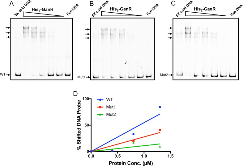
The soil bacterium Bacillus subtilis is often found in association with plants in the rhizosphere. Previously, plant polysaccharides have been shown to stimulate formation of root-associated multicellular communities, or biofilms, in this bacterium, yet the underlying mechanism is not fully understood. A five-gene gan operon (ganSPQAB) in B. subtilis has recently been shown to be involved in utilization of the plant-derived polysaccharide galactan. Despite these findings, molecular details about the regulation of the operon and the role of the operon in biofilm formation remain elusive. In this study, we performed comprehensive genetic analyses on the regulation of the gan operon. We show that this operon is regulated both by a LacI-like transcription repressor (GanR), which directly binds to pairs of inverted DNA repeats in the promoter region of the operon, and by the catabolite control protein A (CcpA). Derepression can be triggered by the presence of the inducer β-1,4-galactobiose, a hydrolysis product of galactan, or in situ when B. subtilis cells are associated with plant roots. In addition to the transcriptional regulation, the encoded ß-galactosidase GanA (by ganA), which hydrolyzes ß-1,4-galactobiose into galactose, is inhibited at the enzymatic level by the catalytic product galactose. Thus, the galactan utilization pathway is under complex regulation involving both positive and negative feedback mechanisms in B. subtilis. We discuss about the biological significance of such complex regulation as well as a hypothesis of biofilm induction by galactan via multiple mechanisms.


Conflict of interest statement

Competing Interests: The authors have declared that no competing interests exist.

Figures

Fig 1
Fig 1. The role of the gan operon in galactan utilization and biofilm formation in B. subtilis.
(A) A working model for the complete galactan utilization pathway in B. subtilis. UDP-Glu and UDP-Gal are the expected end products of galactan utilization carried out by products of the gan and gal genes. UDP-Glu and UDP-Gal are also two essential sugar nucleotide precursors for biosynthesis of exopolysaccharides (EPS) [20]. The Leloir pathway consists of galK, galT, and galE, whose products convert galactose to UDP-Glu [21]. EPS biosynthesis is carried out by enzymes encoded in the espA-O operon, which is indirectly activated by the master regulator Spo0A. Activation of Spo0A by protein phosphorylation in turn depends on multiple sensory histidine kinases including KinB [8]. It is hypothesized in this study that hydrolyzed products of galactan (e.g. galactobiose) can induce kinB expression via the action of GanR (see discussion). (B) Genetic organization of the galactan utilization genes in B. subtilis NCIB3610 and B. licheniformis ATCC8480. Putative promoters and transcription terminators are indicated. Different from B. licheniformis, in the genome of B. subtilis, the galTK genes and the galE gene are separated from the gan operon. In B. subtilis, the gan operon is also only four genes away from the epsA-O operon. Known or proposed functions of the gan and gal genes are as follows: ganSPQ encodes a permease for uptake of galactoligosaccharides; ganA encodes a ß-galactosidase; ganB encodes an endo ß-1,4-galactanse; ganR encodes a transcription repressor; galK encodes a galactokinase; galT for galactose-1-phosphate uridyltransferase; galE for the UDP-galactose-4-epimerase. (C) Development of pellicle biofilms by B. licheniformis ATCC8480, B. subtilis NCIB3610, and B. cereus AR156 in LB supplemented with 0.5% galactan (w/v). LB itself is a biofilm-inert medium for the above strains and used as a control. Images were taken after incubation at 30°C for 3 days. The scale bar represents 0.5 cm.
Fig 2
Fig 2. GanR represses the gan operon and the ganR gene.
(A) Assays of β-galactosidase activities by the reporter strains bearing either PganS-lacZ, or PganR-lacZ, or PganB-lacZ in the wild type strain (blue bars; YC1073, YC1085, and YC1088) and the ganR mutant (red bars; YC1074, YC1086, and YC1089). A deletion mutation in ganA was also introduced into the above strains. Cells were grown in LB shaking broth to OD600 = 1 before harvest and analyses. Assays were done in triplicates and error bars represent standard deviations. (B-C) Assays of ß-galactosidase activities by the wild type reporter strains bearing either PganS-lacZ(YC1073, panel B) or PganR-lacZ(YC1085, panel C). Cells were grown in LB shaking culture over a period of 5.5 hours after inoculation. Both culture densities (red squares, right-hand y-axis) and ß-galactosidase activities of cells (blue diamonds, left-hand y-axis) were measured. Assays were repeated multiple times and representative data was shown here. (D) Assays of ß-galactosidase activities by the PganS-lacZ reporter strains in the wild type background (YC1071), the ΔsinR (YC1091), Δspo0A (YC1092), ΔdegU (YC1248), and ΔccpA (YC1249) mutants. The ganA deletion mutation was not introduced into the above strains. In some mutants, an epsH deletion mutation was also introduced to prevent cell aggregation during shaking growth [45]. Cells were grown in LB shaking culture to OD600 = 1 before harvest and analyses. Error bars represent standard deviations. (E) Display of the promoter regions of ganS and ganR from B. subtilis NCIB3610 and B. licheniformis ATCC8480. The inverted repeats are highlighted in red, the -35 and -10 motifs of the sigma A-dependent promoter are underlined and shown in italic. ATG or GTG start codons of ganS or ganR are highlighted in blue. The cre box for putative CcpA binding sequences in the ganS promoter regions is highlighted in green. The transcriptional start of the ganS gene in B. subtilis was determined in a very recent study [29] and labeled as +1. (F) The consensus DNA motif logo was generated from a multiple sequence alignment of the putative motifs from the selected promoters using WebLogo [31]. The height of each stack, displayed in bits, is representative of the frequency of the nucleotide in the motif.
Fig 3
Fig 3. GanR directly binds to the promoters of ganS and ganR.
(A) Gel mobility shift assays to probe binding of purified His6-GanR to the DNAs containing the promoter sequence of ganS, ganR, or yvaB. Approximately 1 μg (approximately 0.3 μM) of DNA was added to each lane, His6-GanR was added at increasing concentrations from 1, 3, 10, to 30 μM, no protein was added in the control lanes (left-most). Mobility retarded DNA bands were indicated by arrows. (B) The DNA sequence of the ganS promoter in B. subtilis. The -35 and -10 motifs of the sigma A-dependent promoter are highlighted in italic. The putative GanR binding motifs are labeled from Box1 to Box4. (C) A schematic display of site-directed mutagenesis on the putative GanR binding sites in the ganS promoter (mut1, mut2, and mut3). Letters in red are the introduced nucleotide changes in each of the boxes. Mutagenesis in Box4 was avoided due to overlap with the -35 motif of the promoter. (D) Assays of ß-galactosidase activities by the PganS-lacZ reporter strains with either the wild type promoter sequence of ganS, or with various sited-directed mutations shown in (C). Cells were grown in LB shaking culture to OD600 = 1 before harvest and analyses. Error bars represent standard deviations from four independent analyses.
Fig 4
Fig 4. Mutations in inverted DNA repeats in PganS decrease GanR binding.
(A-C) Gel mobility shift assays to determine binding of His6-GanR proteins to the wild type DNA sequence (A), the Mut1 mutagenic sequence (B), and Mut2 mutagenic sequence (C) of the ganS promoter. In all lanes, 16 ng (approximately 5 nM) fluorescent DNA probe was added. His6-GanR proteins were added in a range of concentrations (from 4, 1.3, 0.8, 0.4, to 0.08 μM). In each panel, the right-most lane is the fluorescent probe alone. The left-most lane contains 0.8 μM His6-GanR proteins, 16 ng of fluorescent probe, and 160 ng of unlabeled cold probe for competitive binding. In the upper section in each gel, shifted DNA bands were indicated by arrows. (D) The ratio of shifted versus total DNA was quantified from panels A-C, and graphed to show percent probe shifted versus protein concentration using WT, Mut1, and Mut2 probes.
Fig 5
Fig 5. The gan operon can be induced by galactan, β-1,4-galactobiose, or in situ with plants.
(A) In situ induction of the gan operon by using the PganS-lacZ reporter strain (YC1071). Tomato plant root-associated B. subtilis reporter cells were washed off after 2 days of colonization to tomato plant roots in MG media at 25°C before assays of ß-galactosidase activities. Cells were also applied similarly to the abiotic surface (sterilized bamboo applicator sticks, Fisher Scientific) as a control. (B) Assays of luciferase activities from the PganS-lux reporter strain (YC1146) in the presence of tomato plant root extract (5%, v/v), galactose (0.5%, w/v), glucose (0.5%, w/v), and galactan (0.05%, w/v). Cells were grown in shaking LB broth to OD600 = 1 and luciferase activities were measured using a plate reader (BioTek). (C) Similar assays of luciferase activities from the PganS-lux reporter (YC1146) in the presence of galactose (0.5%, w/v), or ß-1,4-galactobiose (from 0.0025% and 0.02%, w/v). Cells were grown in LB broth with shaking at 37°C in a plate reader and bioluminescence was recorded periodically for 18 hours. The maximal fold induction of the PganS-lux reporter fusion by ß-1,4-galactobiose at hour 16 in was shown here. All assays here were done multiple times and error bars represent standard deviations from those independent assays.
Fig 6
Fig 6. A negative feedback regulation on GanA by its catalytic product galactose.
(A) The wild type strain (3610) and the ganR mutant (YC222S) were streaked out on LB plates supplemented with 40 μg ml-1 X-gal, and without (upper panel) or with (lower panel) galactose (0.5%, w/v). Plates were incubated at 37°C overnight before images were taken. (B-C) Assays of ß-galactosidase activities of protein lysates from cells expressing ganA (panel B, YC222S) or lacZ (panel C, YC1074). Assays were done in the presence of 2.5 mM ONPG and a gradient of galactose (from 2.5 to 20 mM). Error bars represent standard deviations from multiple trials. (D) An overview of complex regulations on galactan utilization involving both (1) a positive feedback mechanism on the transcription of the gan operon by ß-1,4-galactobiose and (2) a negative feedback mechanism at the protein level on GanA by its catalytic product galactose.

References

    1. Lugtenberg B, Kamilova F. Plant-Growth-Promoting Rhizobacteria. Annu Rev Microbiol. 2009;63(1):541–56. doi: 10.1146/annurev.micro.62.081307.162918 . - DOI - PubMed
    1. Earl AM, Losick R, Kolter R. Ecology and genomics of Bacillus subtilis. Trends in Microbiology. 2008;16(6):269–75. doi: 10.1016/j.tim.2008.03.004 - DOI - PMC - PubMed
    1. Ochiai A, Itoh T, Kawamata A, Hashimoto W, Murata K. Plant Cell Wall Degradation by Saprophytic Bacillus subtilis Strains: Gene Clusters Responsible for Rhamnogalacturonan Depolymerization. Applied and Environmental Microbiology. 2007;73(12):3803–13. doi: 10.1128/AEM.00147-07 - DOI - PMC - PubMed
    1. Sakamoto T, Hours A-R, Sakai T. Purification, Characterization, and Production of Two Pectic Transeliminases with Protopectinase Activity from Bacillus subtilis. Bioscience, Biotechnology, and Biochemistry. 1994;58(2):353–8. doi: 10.1271/bbb.58.353 - DOI - PubMed
    1. Soriano M, Diaz P, Pastor FIJ. Pectate lyase C from Bacillus subtilis: a novel endo-cleaving enzyme with activity on highly methylated pectin. Microbiology. 2006;152(3):617–25. doi: 10.1099/mic.0.28562–0 - DOI - PubMed

MeSH terms

LinkOut - more resources