. 2017 Jun 15;13(6):e1006427.
doi: 10.1371/journal.ppat.1006427. eCollection 2017 Jun.

Wolbachia elevates host methyltransferase expression to block an RNA virus early during infection

Affiliations

Wolbachia elevates host methyltransferase expression to block an RNA virus early during infection

Tamanash Bhattacharya et al. PLoS Pathog. .

Abstract

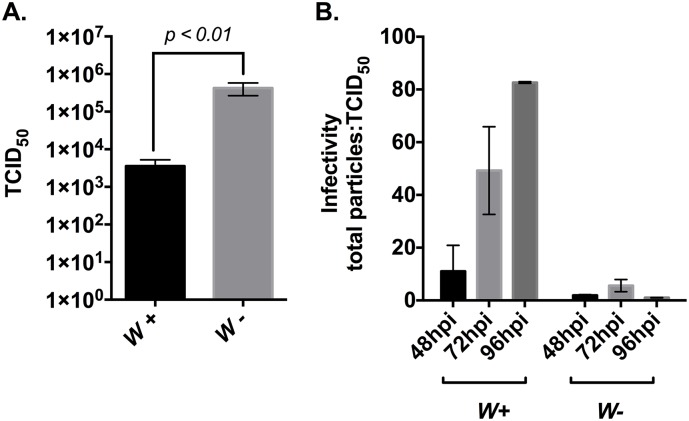
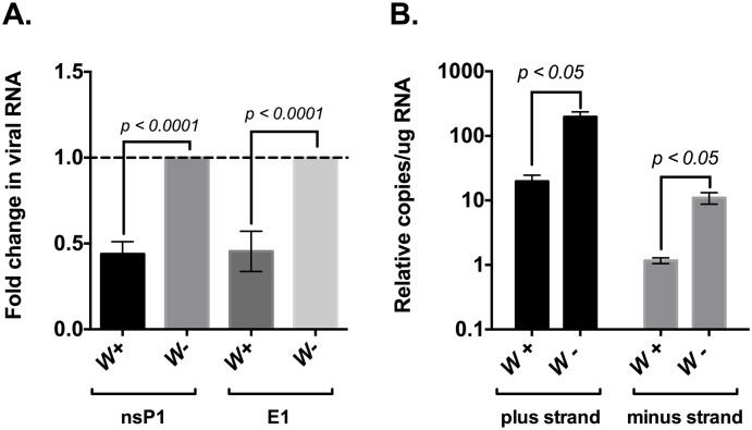
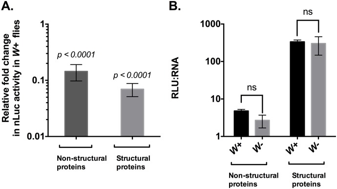
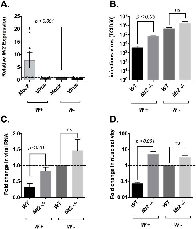
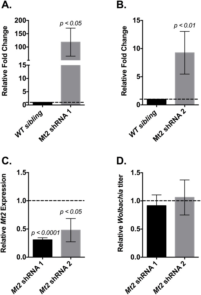
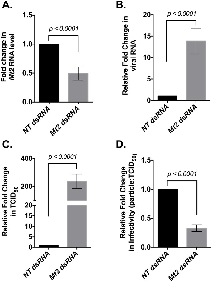
Wolbachia pipientis is an intracellular endosymbiont known to confer host resistance against RNA viruses in insects. However, the causal mechanism underlying this antiviral defense remains poorly understood. To this end, we have established a robust arthropod model system to study the tripartite interaction involving Sindbis virus and Wolbachia strain wMel within its native host, Drosophila melanogaster. By leveraging the power of Drosophila genetics and a parallel, highly tractable D. melanogaster derived JW18 cell culture system, we determined that in addition to reducing infectious virus production, Wolbachia negatively influences Sindbis virus particle infectivity. This is further accompanied by reductions in viral transcript and protein levels. Interestingly, unchanged ratio of proteins to viral RNA copies suggest that Wolbachia likely does not influence the translational efficiency of viral transcripts. Additionally, expression analyses of candidate host genes revealed D. melanogaster methyltransferase gene Mt2 as an induced host factor in the presence of Wolbachia. Further characterization of viral resistance in Wolbachia-infected flies lacking functional Mt2 revealed partial recovery of virus titer relative to wild-type, accompanied by complete restoration of viral RNA and protein levels, suggesting that Mt2 acts at the stage of viral genome replication. Finally, knockdown of Mt2 in Wolbachia uninfected JW18 cells resulted in increased virus infectivity, thus demonstrating its previously unknown role as an antiviral factor against Sindbis virus. In conclusion, our findings provide evidence supporting the role of Wolbachia-modulated host factors towards RNA virus resistance in arthropods, alongside establishing Mt2's novel antiviral function against Sindbis virus in D. melanogaster.


Conflict of interest statement

The authors have declared that no competing interests exist.

Figures

Fig 1
Fig 1. Effect of Wolbachia on SINV infectivity.
(A) Presence of Wolbachia is correlated with reduced virus titer in D. melanogaster flies. Systemic viral infection was established in Wolbachia-infected (left) and Wolbachia-free (right) flies as described in Materials and Methods. Infection was allowed to last for 48-hours before whole fly tissues were harvested and assayed for the presence of infectious virus using end-point-dilution assay in BHK-21 cells. Numbers along the y-axis represent the dilutions that resulted in at least 50 percent cell death (TCID50). Data are mean values for three independent experiments. P values were calculated using Mann-Whitney U test (p = 0.0050) (B) Presence of Wolbachia in D. melanogaster derived JW18 cells lead to reduced virus particle infectivity over time. Wolbachia-infected (left) and Wolbachia-free (right) JW18 cells were infected with SINV at an MOI of 100 and media was collected at 48, 72 and 96 hours post infection. Samples were subsequently assayed for infectious virus using end-point-dilution assay and for particle numbers by qRT-PCR quantification of viral genome copies present in the media. Higher total particle: TCID50 numbers are indicative of lower virus particle infectivity. Values reported are the mean particle-to-TCID50 ratios of two independent replicates. All error bars represent standard error of mean (SEM).
Fig 2
Fig 2. Wolbachia reduces SINV RNA synthesis.
(A) Systemic virus infection was established in Wolbachia–infected (left) and Wolbachia–free (right) flies as described in Materials and Methods. Total RNA was isolated from fly tissue homogenates 48 hours post infection and assayed for fold change in RNA synthesis using qRT-PCR. Values reported are the mean of three independent biological replicates. P values were calculated using Mann-Whitney U test (nsP1: p < 0.0001, E1: p < 0.0001) (B) Absolute quantification of viral plus strand (left) and minus strand (right) RNA species observed during SINV infection in Wolbachia–infected and Wolbachia–free JW18 cells. Cells were infected with SINV at an MOI of 100 that lasted 96 hours, followed by extraction of total cellular RNA. Quantification of SINV RNA species was performed using qRT-PCR as described in Materials and Methods. Values were calculated using standard curves. P values were calculated using Unpaired t-test with Welch’s correction (plus strand: p = 0.043, t = 4.521, df = 2.063, minus strand: p = 0.046, t = 4.457, df = 2.012). All error bars represent standard error of mean (SEM).
Fig 3
Fig 3. Viral protein synthesis is reduced in the presence of Wolbachia in flies.
(A) Wolbachia–infected and Wolbachia–free flies were each infected with either SINV-nsP3-nLuc (left) or SINV cap-nLuc (right) viruses as described in Materials and Methods. Infection was allowed to last for 48 hours and fly tissues were harvested and assayed for luciferase activity to determine viral nonstructural (left) and structural (right) protein levels respectively. Reported values are relative to nLuc activity observed in Wolbachia–free (W-) flies, each set at a value of 1. Values represented the mean of three independent biological replicates. P values were calculated using Mann-Whitney U test (non-structural proteins: p < 0.0001, structural proteins: p < 0.0001) (B) Translation efficiency (RLU: RNA) of SINV non-structural (left) and structural (right) polyproteins was calculated as the ratio of total protein (shown above as the net luciferase output or RLU) to absolute copies of viral genomic and subgenomic transcripts, respectively. Absolute quantification of viral genomic and subgenomic RNA species was performed using qRT-PCR as described before. Values represented the mean of three independent biological replicates. P values were calculated using Mann-Whitney U test (non-structural proteins: p = 0.20, structural proteins: p > 0.99). All error bars represent standard error of mean (SEM).
Fig 4
Fig 4. Wolbachia induced SINV resistance is dependent on host methyltransferase gene Mt2.
(A) Mt2 expression profile in Wolbachia–infected (left)and Wolbachia–free (right) flies either in the absence or presence of SINV infection. Virus infection was established as described in Materials and Methods. Infection was allowed to last for 48 hours followed by quantification of Mt2 expression via qRT-PCR. P values were calculated using Mann-Whitney U test (p < 0.001) (B) Quantification of infectious virus produced during SINV infection in Wolbachia–infected (left)and Wolbachia–free (right) flies that are either wild-type (WT) or lacking functional Mt2 gene (Mt2 -/-). Flies were challenged with SINV for 48 hours before whole fly tissues were harvested and assayed for the presence of infectious virus using end-point-dilution assay in BHK-21 cells. Values represent the mean of three independent biological replicates. P values were calculated using Mann-Whitney U tests between genotypes (p < 0.05) (C) Effect of Mt2 loss on viral RNA synthesis in the presence of Wolbachia. SINV RNA was quantified using qRT-PCR by probing against the viral E1 gene. Values reported above are relative to viral RNA levels observed in Wolbachia–free wild-type flies. Values represent the mean of three independent biological replicates. P values were calculated using Mann-Whitney U tests between genotypes (W+: p < 0.01, W-: p = 0.71) (D) Wolbachia–infected (left)and Wolbachia–free (right) wild-type or Mt2 -/- flies were challenged with SINV cap-nLuc virus. After 48 hours post infection, fly tissues were harvested and assayed for luciferase activity to determine viral structural protein levels. Reported values are relative to nLuc activity observed in wild-type Wolbachia–free (W-) flies, set at 1. Values represent the mean of three independent biological replicates. P values were calculated using Mann-Whitney U tests between genotypes (W+: p < 0.001, W-: p = 0.224). All error bars represent standard error of mean (SEM).
Fig 5
Fig 5. Effect of TRiP knockdown of Mt2 expression in Wolbachia infected flies.
Mt2 expression was knocked down in Wolbachia infected transgenic RNAi fly stocks 38224 (Mt2 shRNA 1) and 42906 (Mt2 shRNA 2) by driving Mt2 shRNA expression via chromosome II Act5C-Gal4 driver (y1 w*; P{Act5C-GAL4}25FO1/CyO) as described in Materials and Methods. For each set of crosses, siblings lacking the expression of Mt2 targeting shRNA were used as the wild-type controls. (A, B) Flies were challenged with SINV as described previously whereby infection was allowed to last for 48 hours before whole fly tissues were collected and assayed for infectious virus using end-point-dilution assay on BHK-21 cells. Values represent the mean of six independent biological replicates. (C) Quantitative analyses of Mt2 expression in TRiP mutant flies relative to their respective wild-type sibling controls (set at 1) was performed using qRT-PCR on total RNA extracted from fly tissues. Values for each column represent the mean of 6 and 3 independent biological replicates, respectively. P values were calculated using Mann-Whitney U tests (Mt2 shRNA 1: p < 0.0001, Mt2 shRNA 2: p = 0.0361). (D) Wolbachia titer was quantified using qPCR. Reported values are relative to respective wild-type sibling controls (set at 1) and are represented as the mean of two independent biological replicates. All error bars represent standard error of mean (SEM).
Fig 6
Fig 6. Mt2 antiviral activity does not depend on the Wolbachia infection status of the host.
Mt2 expression was knocked down in D. melanogaster derived Wolbachia–free JW18 cells using dsRNA against the host methyltransferase gene. Approximately 48-hours post-transfection, cells were challenged with SINV at an MOI of 100 as described in Materials and Methods. Infection was allowed to last for 48 hours before harvesting the cells and media. Total cellular RNA was extracted from cells as described in Materials and Methods and assayed for host Mt2 (A) and viral E1 (B) gene expression. Values represent the mean of five independent biological replicates. P values were calculated using Mann-Whitney U tests (Mt2: p < 0.0001, SINV E1: p < 0.0001). (C) Media was collected 48 hours post infection and infectious virus particle was quantified as before, using end-point-dilution assay on BHK-21cells. Values represent the mean of five independent biological replicates. P values were calculated using Mann-Whitney U tests (TCID50: p < 0.0001). (D) Infectivity of virus particles produced was determined as described earlier, whereby total number of virus particles were quantified using qRT-PCR. As before, higher total particle: TCID50 numbers indicate poorer virus particle infectivity and relative reduction in particle: TCID50 ratio represents an overall increase in infectivity. Values represent the mean of five independent biological replicates. P values were calculated using Mann-Whitney U tests (particle: TCID50: p < 0.0001). All reported values are relative to non-targeting, control dsRNA treatments set at 1. All error bars represent standard error of mean (SEM).

Similar articles

Cited by

References

    1. Bennett GM, Moran NA. Heritable symbiosis: The advantages and perils of an evolutionary rabbit hole. Proceedings of the National Academy of Sciences. 2015. August 18;112(33):10169–76. - PMC - PubMed
    1. Werren JH, Baldo L, Clark ME. Wolbachia: master manipulators of invertebrate biology. Nature Reviews Microbiology. 2008. October 1;6(10):741–51. 10.1038/nrmicro1969 - DOI - PubMed
    1. Zug R, Hammerstein P. Still a host of hosts for Wolbachia: analysis of recent data suggests that 40% of terrestrial arthropod species are infected. PloS one. 2012. June 7;7(6):e38544 10.1371/journal.pone.0038544 - DOI - PMC - PubMed
    1. Hoffmann AA, Hercus M, Dagher H. Population dynamics of the Wolbachia infection causing cytoplasmic incompatibility in Drosophila melanogaster. Genetics. 1998. January 1;148(1):221–31. - PMC - PubMed
    1. Teixeira L, Ferreira Á, Ashburner M. The bacterial symbiont Wolbachia induces resistance to RNA viral infections in Drosophila melanogaster. PLoS Biol. 2008. December 23;6(12):e1000002. - PMC - PubMed

MeSH terms

Substances

LinkOut - more resources