. 2017 Jun 15;13(6):e1006404.
doi: 10.1371/journal.ppat.1006404. eCollection 2017 Jun.

Hypoxia-inducible factor-1α plays roles in Epstein-Barr virus's natural life cycle and tumorigenesis by inducing lytic infection through direct binding to the immediate-early BZLF1 gene promoter

Affiliations

Hypoxia-inducible factor-1α plays roles in Epstein-Barr virus's natural life cycle and tumorigenesis by inducing lytic infection through direct binding to the immediate-early BZLF1 gene promoter

Richard J Kraus et al. PLoS Pathog. .

Abstract

When confronted with poor oxygenation, cells adapt by activating survival signaling pathways, including the oxygen-sensitive transcriptional regulators called hypoxia-inducible factor alphas (HIF-αs). We report here that HIF-1α also regulates the life cycle of Epstein-Barr virus (EBV). Incubation of EBV-positive gastric carcinoma AGS-Akata and SNU-719 and Burkitt lymphoma Sal and KemIII cell lines with a prolyl hydroxylase inhibitor, L-mimosine or deferoxamine, or the NEDDylation inhibitor MLN4924 promoted rapid and sustained accumulation of both HIF-1α and lytic EBV antigens. ShRNA knockdown of HIF-1α significantly reduced deferoxamine-mediated lytic reactivation. HIF-1α directly bound the promoter of the EBV primary latent-lytic switch BZLF1 gene, Zp, activating transcription via a consensus hypoxia-response element (HRE) located at nt -83 through -76 relative to the transcription initiation site. HIF-1α did not activate transcription from the other EBV immediate-early gene, BRLF1. Importantly, expression of HIF-1α induced EBV lytic-gene expression in cells harboring wild-type EBV, but not in cells infected with variants containing base-pair substitution mutations within this HRE. Human oral keratinocyte (NOK) and gingival epithelial (hGET) cells induced to differentiate by incubation with either methyl cellulose or growth in organotypic culture accumulated both HIF-1α and Blimp-1α, another cellular factor implicated in lytic reactivation. HIF-1α activity also accumulated along with Blimp-1α during B-cell differentiation into plasma cells. Furthermore, most BZLF1-expressing cells observed in lymphomas induced by EBV in NSG mice with a humanized immune system were located distal to blood vessels in hypoxic regions of the tumors. Thus, we conclude that HIF-1α plays central roles in both EBV's natural life cycle and EBV-associated tumorigenesis. We propose that drugs that induce HIF-1α protein accumulation are good candidates for development of a lytic-induction therapy for treating some EBV-associated malignancies.


Conflict of interest statement

The authors have declared that no competing interests exist.

Figures

Fig 1
Fig 1. Hypoxia mimics induce accumulation of lytic EBV proteins in some EBV+ cell lines.
(A)) Immunoblots showing accumulation of HIF-1α and Zta after incubation of Sal cells with the indicated concentrations of L-Mimosine for 24 h. (B) Immunoblots showing relative levels of HIF-1α and the indicated lytic EBV antigens present in AGS-Akata and SNU-719 cells 24 h after addition to the medium of DFO (200 μM; lanes 2 and 5), MLN4924 (5 μM; lanes 3 and 6), or the diluent vehicle (lanes 1 and 4). (C) Immunoblots showing accumulation of HIF-1α and the EBV-encoded proteins Zta, EAD, and VCA/p18 after incubation of AGS-Akata cells with (+) or without (-) 200 μM DFO for 24 h followed by immediate harvesting (lanes 1 and 2) or incubation for an additional 24 h in the absence of DFO prior to harvesting (lanes 3 and 4). (D) Immunoblots showing the relative levels of accumulation of the indicated proteins in several cells lines incubated in the presence (+) or absence (-) of 200 μM DFO for 24 h. (E) Immunoblots comparing accumulation of HIF-1α and Zta in Sal versus KemIII cells after incubation as described in panel D. Whole-cell extracts were prepared, processed, and probed for the indicated proteins. GAPDH served as a loading control. Data shown are representative of numerous independent sets of experiments.
Fig 2
Fig 2. Dual immunofluorescence staining indicates DFO efficiently induces synthesis of Zta protein in a subset of HIF-1α-expressing EBV-positive cells.
AGS-Akata cells grown on cover slips were incubated for 24 h in the absence (-) or presence (+) of 200 μM DFO prior to fixing and processing for co-detection by IFS of the proteins Zta (green) and HIF-1α (red). DFO-treated cells were independently probed with the green-conjugated secondary antibody absent primary antibodies to control for background GFP encoded by the virus.
Fig 3
Fig 3. HIF-1α addition is sufficient to induce EBV reactivation and necessary for efficient induction by DFO.
(A) Immunoblots showing addition of HIF-1α is sufficient to induce lytic EBV reactivation. AGS-Akata cells grown in 10-cm dishes were transfected with 1 μg each of pHA-HIF-1α P402A/P564A-pcDNA3 plus pHIF-β (+) or 2 μg of their empty vector, pcDNA3, as a control (-) and incubated for 48 h prior to preparation of whole-cell extracts. Data are representative of numerous independent experiments. (B) Immunoblots showing knockdown of HIF-1α inhibits DFO-induced synthesis of EBV lytic antigens. Lanes 1–6, AGS-Akata cells maintained in 10-cm dishes were co-transfected with 0.8 μg of each of five lentiviruses that express different shRNAs targeting HIF-1α (lanes 5–6) or 4 μg of a lentivirus that expresses the non-targeting shRNA cntl. #1 or cntl. #2 (lanes 1–2 and lanes 3–4, respectively). Two days later, the cells were incubated in the absence (-) or presence (+) of 200 μM DFO for 24 h prior to harvesting and preparation of whole-cell extracts. Lanes 7–10, Sal cells were infected with the indicated packaged lentiviruses; three days later, the cells were incubated in the absence (-) or presence (+) of 200 μM DFO for 24 h and processed likewise. Data are representative of two independent experiments. GADPH served as a loading control.
Fig 4
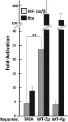
Fig 4. HIF-1α induces transcriptional activation from Zp, but not Rp.
293T cells maintained in 24-well plates were co-transfected with (i) 200 ng DNA of a pGL3-Basic luciferase reporter containing the nt -30 to +30 region of the HSV TK gene (pTATA-luc) as a control, the nt -221 to +30 region of Zp (pWTZp-luc), or the nt -1069 to +38 region of Rp (pWTRp-luc), and (ii) pHA-HIF-1αP402A/P564A-pcDNA3 plus pHIF-1β (40 ng each), pcDNA3-BRLF1 (30 ng) as a positive control, or pcDNA3 (80 ng) as a negative control. Cells were harvested 48 h later, and luciferase activities were determined. Data obtained with each reporter were normalized to the value obtained when co-transfected with pcDNA3; they are averages from three independent experiments each performed in triplicate; error bars indicate standard errors of the mean. **, p < 0.01.
Fig 5
Fig 5. Zp contains a transcriptionally functional HRE activated by both HIF-1α and HIF-2α.
(A) In silico identification of a consensus HRE located from nt -83 through -76 relative to the transcription initiation site of Zp. Shown here is a schematic indicating cis-acting regulatory elements (denoted by rectangles) along with their trans-acting factors where known. SBE, SMAD-binding element. (B) Schematic indicating the sequence alterations present in the base pair substitution mutants, M1-M4 and ZIIRM, analyzed here. The lines above the sequence indicate the locations of the HRE, ZIIR, and ZII elements. (C, D) Both HIF-1α- and HIF-2α-dependent activation of transcription from Zp map to the putative HRE. 293T cells maintained in 24-well plates were co-transfected with (i) 200 ng of pTATA-luc as a control, pWTZp-luc, or the base pair substitution variants of pWTZp-luc depicted in Fig. 5B, and (ii) pHA-HIF-1αP402A/P564A-pcDNA3 (panel C) or pHA-HIF-2αP405A/P531A-pcDNA3 (panel D) plus pHIF-1β (40 ng each) or 80 ng pcDNA3. Cells were harvested 48 h later, and luciferase activities were determined as described in the legend to Fig 4. Data are averages from three or more independent experiments each performed in triplicate. **, p < 0.01.
Fig 6
Fig 6. HIF-1α sequence-specifically binds the nt -80 region Zp HRE.
(A) EMSA showing HIF-1α binding to a radiolabeled, double-stranded oligonucleotide that contains the consensus HRE WT sequence shown in panel C. Approximately 30 μg protein obtained from a nuclear extract prepared from CoCl2–treated AGS cells was pre-incubated with 1 μg anti-HIF-1α polyclonal antibody (lane 2) or 1 μg anti-IgG antibody as a control (lane 1) prior to addition of the probe DNA and electrophoresis. (B) Competition EMSA showing sequence-specific binding of HIF-1α to the Zp HRE. Assays were performed by pre-incubation of the reaction mixture with the indicated unlabeled, double-stranded competitor oligonucleotides at the indicated amounts prior to addition of the radiolabeled probe and electrophoresis. (C) Sequences of the oligonucleotides used as probe (HRE WT) and competitors. Mutated bases indicated by italicized font. Boxes, HREs.
Fig 7
Fig 7. HIF-1α binds to Zp in context of whole EBV genomes.
(A) Immunoblots showing relative levels of HIF-1α and Zta present in Sal and SNU-719 cells incubated for 24 h with (+) or without (-) 200 μM DFO prior to processing for ChIP analysis. (B, C) Quantitative-PCR analyses of the chromatin obtained from the cells in panel A following precipitation with anti-HIF-1α or anti-IgG antibodies. The primer pairs spanned Zp versus a sequence located 4.8-kbp upstream of Zp as a negative control (neg. cntl.). Data presented are average Ct values of two independent experiments each performed in triplicate; error bars indicate standard deviations. **, p < 0.01; ***, p < 0.001.
Fig 8
Fig 8. HIF-α-induced lytic reactivation of EBV requires the Zp HRE.
Even-numbered lanes (+), the indicated WT- and HRE mutant-infected 293T cell lines grown in 6-well plates were transfected with: (A) 0.5 μg each of plasmids expressing HIF-1β and the oxygen-insensitive variant of HIF-1α; (B) 50 ng of the Zta-expressing plasmid, pCMV-BZLF1; (C) 100 ng of the Rta-expressing plasmid, pRTS15; or (D) 0.5 μg each of plasmids expressing HIF-1β and the oxygen-insensitive variant of HIF-2α. Whole-cell extracts were prepared 72 h later and analyzed by immunoblotting for the indicated proteins. Correspondingly similar amounts of pcDNA3 DNA were transfected in parallel in the odd-numbered lanes (-) as controls.
Fig 9
Fig 9. Differentiation of memory B cells into plasmablasts induces HIF-1α activity.
Changes in RNA levels during differentiation of primary human B cells into plasma cells of some cellular genes (VEGFA, PDK1) whose expression is known to be activated by HIF-1α and some other genes whose products (ZEB1, ZEB2, Blimp-1α, XBP-1s) are known to contribute to regulation of BZLF1 gene expression. Relative levels of these RNAs were determined by extraction of data from five sets of microarray analyses of mRNA purified from cells harvested at the eight indicated stages of B-cell differentiation. Note large differences in scales shown for y-axes.
Fig 10
Fig 10. Differentiation of epithelial cells induces accumulation of HIF-1α.
Immunoblots showing changes with time in HIF-1α protein levels following induction of differentiation of (A) NOK and (B) NOK-Akata cells by suspension in methylcellulose (1.6% in K-SFM) for the times indicated. (C) Immunoblots showing HIF-1α accumulation in hGET cells following induction of differentiation by suspension in methyl cellulose (1.6% in K-SFM for 48 h; lane 2). As controls, the cells for lanes 1, 3, and 4 were incubated in parallel in K-SFM without MC. For lane 4, 50 μM DFO was added to the cells 24 h prior to harvest as a control for HIF-1α stabilization in the absence of MC-induced differentiation. (D) Immunoblots showing HIF-1α accumulation in NOK (clone #3) cells following induction of differentiation by growth in organotypic culture for 11 days at an air-liquid interface. Lanes 2–4 contain protein extracts from three different rafts. Lane 1 contains protein extract from NOK (clone #1) cells maintained in an undifferentiated state as a monolayer grown in K-SFM. Whole-cell extracts were prepared and analyzed for the indicated proteins.
Fig 11
Fig 11. Most Zta+ cells present in B-cell lymphomas induced by EBV in mice with a humanized immune system reside distal to blood vessels.
NSG mice were inoculated i.p. with human cord blood that had been infected 1.5 h earlier with the M81 strain of EBV. Thirty-three days later, the mice were sacrificed, and the tumors were flash frozen, sectioned, and processed by IFS for the indicated proteins. (A) Sections co-stained for EBNA2 (green) and CD31 (red). (B) Sections co-stained for Zta (green) and CD31 (red). (C) Sections co-stained for Zta (green) and Hypoxyprobe (red). All sections were counterstained with DAPI (blue). These sections are representative of data observed with over two dozen EBV+ tumors obtained in several experiments performed with blood from different donors. Arrows indicate locations of some of the blood vessels based upon cross-reactivation with CD31 antibody.
Fig 12
Fig 12. Histogram showing distributions of distances of EBNA2+ versus Zta+ cells from the nearest blood vessel (i.e., a CD31+ cell).
Distances for 263 EBNA2+ and 263 Zta+ cells were measured from stained sections similar to the ones shown in Fig 11 and S3 Fig.
Fig 13
Fig 13. Model for how differentiation of epithelial and B cells may induce reactivation of EBV into lytic infection and influence ability to be infected by EBV via altering levels of cellular transcription factors known to contribute to repression and activation of BZLF1 and BRLF1 gene expression.
See text for details.

Similar articles

Cited by

References

    1. Longnecker RM, Kieff E, Cohen JI. Epstein-Barr virus Fields Virology. 6th edition Lippincott, Williams, and Willams; 2013.
    1. Thorley-Lawson DA. EBV Persistence—Introducing the Virus. Curr Top Microbiol Immunol. 2015;390: 151–209. doi: 10.1007/978-3-319-22822-8_8 - DOI - PMC - PubMed
    1. Kang M-S, Kieff E. Epstein-Barr virus latent genes. Exp Mol Med. 2015;47: e131 doi: 10.1038/emm.2014.84 - DOI - PMC - PubMed
    1. Kutok JL, Wang F. Spectrum of Epstein-Barr virus-associated diseases. Annu Rev Pathol. 2006;1: 375–404. doi: 10.1146/annurev.pathol.1.110304.100209 - DOI - PubMed
    1. Rickinson AB. Co-infections, inflammation and oncogenesis: future directions for EBV research. Semin Cancer Biol. 2014;26: 99–115. doi: 10.1016/j.semcancer.2014.04.004 - DOI - PubMed

MeSH terms

Substances