. 2018 Mar;79(3):1241-1250.
doi: 10.1002/mrm.26788. Epub 2017 Jun 15.

Improved localization, spectral quality, and repeatability with advanced MRS methodology in the clinical setting

Affiliations

Improved localization, spectral quality, and repeatability with advanced MRS methodology in the clinical setting

Dinesh K Deelchand et al. Magn Reson Med. 2018 Mar.

Abstract

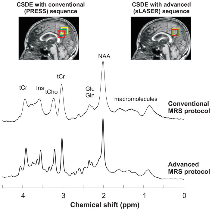
Purpose: To investigate the utility of an advanced magnetic resonance spectroscopy (MRS) protocol in the clinical setting, and to compare the localization accuracy, spectral quality, and quantification repeatability between this advanced and the conventional vendor-provided MRS protocol on a clinical 3T platform.

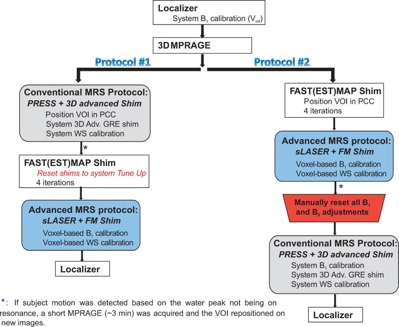
Methods: Proton spectra were measured from the posterior cingulate cortices in 30 healthy elderly subjects by clinical MR technologists using a vendor-provided (point resolved spectroscopy with advanced 3D gradient-echo B0 shimming) and an advanced (semi-LASER with FAST(EST)MAP shimming) protocol, in random order. Spectra were quantified with LCModel using standard pipelines for the clinical and research settings, respectively.

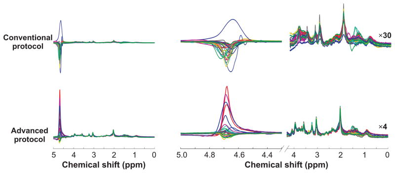
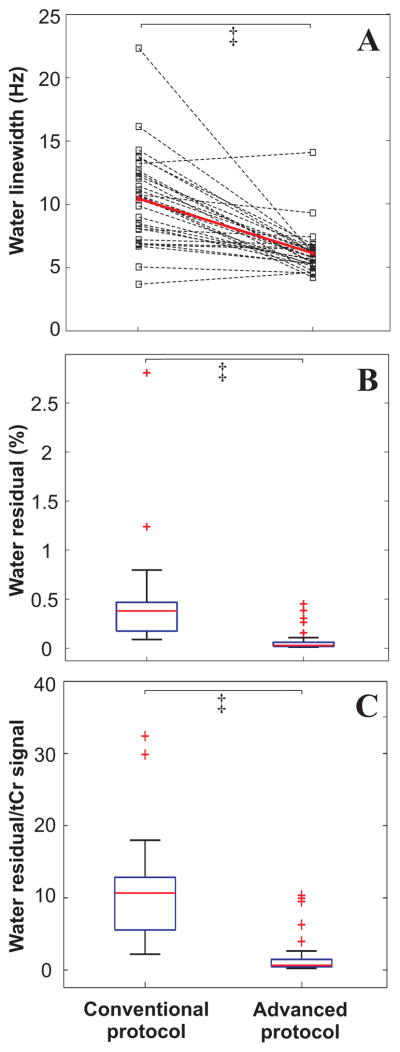
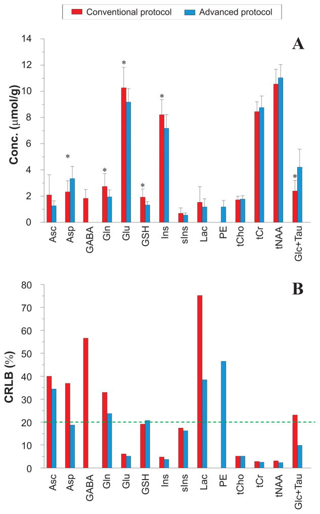
Results: The advanced protocol outperformed the vendor-provided protocol in localization accuracy (chemical-shift-displacement error: 2.0%/ppm, semi-LASER versus 11.6%/ppm, point resolved spectroscopy), spectral quality (water linewidth: 6.1 ± 1.8 Hz, FAST(EST)MAP versus 10.5 ± 3.7 Hz, 3D gradient echo; P < 7e-6; residual water: 0.08 ± 0.12%, VAPOR versus 0.45 ± 0.50%, WET; P < 2e-5) and within-session repeatability of metabolite concentrations, particularly of low signal-to-noise ratio data with two to eight averages (test-retest coefficients of variance of metabolite concentrations, P < 0.01). Concentrations of J-coupled metabolites such as γ-aminobutyric acid and glutamate were biased when using the default pipeline with simulated macromolecules.

Conclusions: The quality of MRS data can be improved using advanced acquisition and analysis protocols on standard 3T hardware in the clinical setting, which can facilitate robust applications in central nervous system diseases. Magn Reson Med 79:1241-1250, 2018. © 2017 International Society for Magnetic Resonance in Medicine.

Keywords: 3T; FAST(EST)MAP; PRESS; chemical shift displacement; linewidth; sLASER.


Figures

Figure 1
Figure 1
Randomized MRS protocols (that consist of the conventional and advanced acquisition schemes in alternate order) carried out by three rotating neuroradiology MR technologists at 3 T. PCC: posterior cingulate cortex; WS: water suppression; FM: FAST(EST)MAP. A localizer was acquired at the end of the study to verify that the subject has not changed their position in the scanner. The advanced MRS protocol was slightly longer by ~2 mins due to the extra steps of adjusting the B1 levels for the 90° and WS pulses.
Figure 2
Figure 2
PRESS (TR/TE = 5000/30 ms) and sLASER (TR/TE = 5000/28 ms) spectra (64 averages) acquired at 3T from one subject using the conventional and advanced MRS protocols, respectively. Displayed spectra were processed with 3.8 Hz Gaussian weighting. Although similar spectral patterns were observed between the different protocols, broader spectral linewidth was obtained with the Adv. 3D shim compared to FM shim. CSDE for the selected VOI (green) observed with PRESS and sLASER are shown for two resonances with a chemical shift difference of ±1.25 ppm relative to transmitter offset (red and yellow boxes) on T1-weighted images. Measured linewidth for tCr-CH3 was 12 and 4 Hz with the conventional and advanced protocols respectively. The broader linewidth observed with the conventional protocol was due to B0 shimming technique and small shot-to-shot frequency shifts.
Figure 3
Figure 3
Overlaid spectra (exponentially line-broadened by 0.5 Hz for display purpose only) acquired from 30 subjects using the conventional and advanced protocols. The upfield region (0 to 4.1 ppm in the right panel) was appropriately scaled relative to the water residual peak (30-fold multiplication for PRESS spectra, 4-fold for sLASER spectra) to show the reproducibility of spectra between subjects.
Figure 4
Figure 4
Performance of voxel based B0 shimming routines (Adv. 3D versus FM) and water suppression schemes (WET versus VAPOR). FM shim resulted in narrower spectral linewidth than the Adv. 3D protocol (A) and better water suppression was achieved with VAPOR than the WET scheme (B and C). ‡ P<2.5e-5 (Wilcoxon test).
Figure 5
Figure 5
Mean metabolite concentrations (A) and CRLBs (B) obtained with the conventional and advanced MRS acquisition and analysis protocols. Error bars represent intersubject standard deviations and the green dotted line represents a mean CRLB cut-off of 20%. * P<0.01 (two-tailed, paired Student’s t-test) between conventional versus advanced protocol. Only differences at the level of P<0.01 are shown due to multiple comparisons. Ascorbate (Asc), aspartate (Asp), γ-aminobutyric acid (GABA), glutamine (Gln), glutamate (Glu), glutathione (GSH), myo-inositol (Ins), scyllo-inositol (sIns), lactate (Lac), phosphorylethanolamine (PE), glycerophosphorylcholine + phosphorylcholine (tCho), creatine + phosphocreatine (tCr), N-acetylaspartate + N-acetylaspartylglutamate (tNAA), glucose + taurine (Glc+Tau).
Figure 6
Figure 6
Mean repeatability CVs for metabolites with mean CRLB ≤20% as determined from Figure 5. Error bars represent intersubject standard deviations. * P<0.01 (two-tailed, paired Student’s t-test) between conventional versus advanced protocols.

References

    1. Öz G, Alger J, Barker P, Bartha R, Bizzi A, Boesch C, Bolan P, Brindle K, Cudalbu C, Dincer A, Dydak U, Emir U, Frahm J, Gonzalez R, Gruber S, Gruetter R, Gupta R, Heerschap A, Henning A, Hetherington H, Howe F, Huppi P, Hurd R, Kantarci K, Klomp D, Kreis R, Kruiskamp M, Leach M, Lin A, Luijten P, Marjanska M, Maudsley A, Meyerhoff D, Mountford C, Nelson S, Ozduman K, Necmettin P, Pan J, Peet A, Poptani H, Posse S, Pouwels P, Ratai E, Ross B, Scheenen T, Schuster C, Soher B, Tkac I, Vigneron D, Kauppinen R The MRS Consensus Group. Clinical Proton MR Spectroscopy in Central Nervous System Disorders. Radiology. 2014;270(3):658–679. - PMC - PubMed
    1. Terpstra M, Cheong I, Lyu T, Deelchand DK, Emir UE, Bednařík P, Eberly LE, Öz G. Test-retest reproducibility of neurochemical profiles with short-echo, single-voxel MR spectroscopy at 3T and 7T. Magn Reson Med. 2016;76(4):1083–1091. - PMC - PubMed
    1. Deelchand DK, Adanyeguh IM, Emir UE, Nguyen T-M, Valabregue R, Henry P-G, Mochel F, Öz G. Two-site reproducibility of cerebellar and brainstem neurochemical profiles with short-echo, single-voxel MRS at 3T. Magn Reson Med. 2015;73(5):1718–1725. - PMC - PubMed
    1. Frahm J, Merboldt K-D, Hanicke W. Localized proton spectroscopy using stimulated echoes. J Magn Reson. 1987;72(3):502–508. - PubMed
    1. Bottomley PA. Spatial Localization in NMR Spectroscopy in Vivo. Annals of the New York Academy of Sciences. 1987;508(1):333–348. - PubMed

Publication types