. 2017 Jun 14;21(6):671-681.e4.
doi: 10.1016/j.chom.2017.05.009.

IL-22 Upregulates Epithelial Claudin-2 to Drive Diarrhea and Enteric Pathogen Clearance

Affiliations

IL-22 Upregulates Epithelial Claudin-2 to Drive Diarrhea and Enteric Pathogen Clearance

Pei-Yun Tsai et al. Cell Host Microbe. .

Abstract

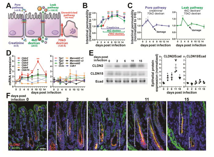
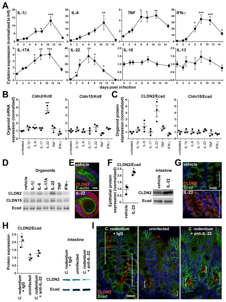
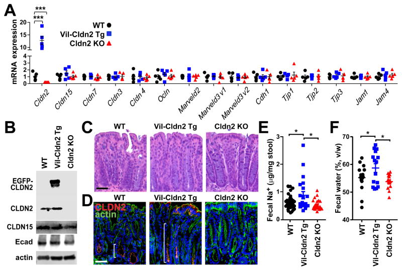
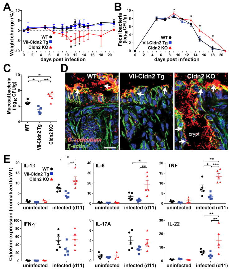
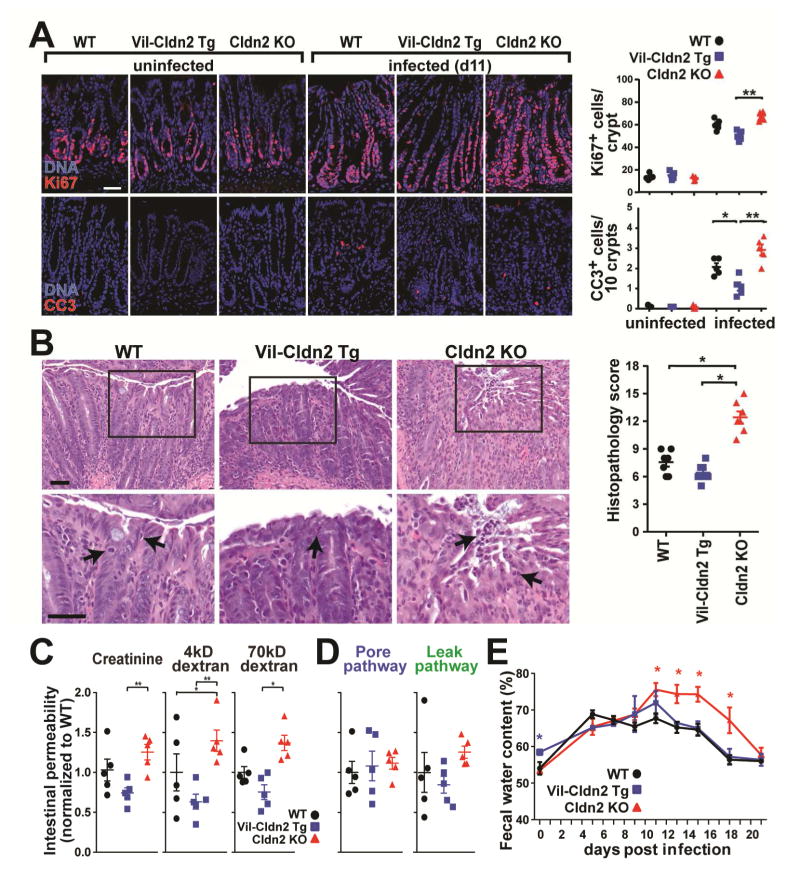
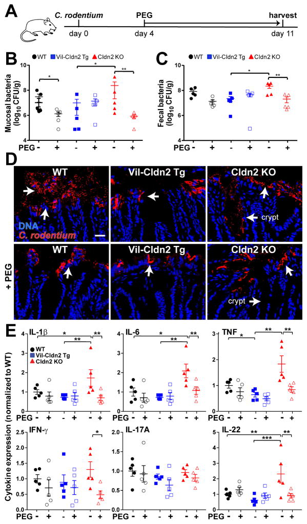
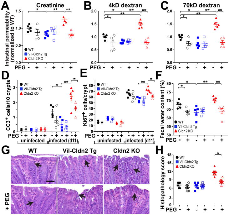
Diarrhea is a host response to enteric pathogens, but its impact on pathogenesis remains poorly defined. By infecting mice with the attaching and effacing bacteria Citrobacter rodentium, we defined the mechanisms and contributions of diarrhea and intestinal barrier loss to host defense. Increased permeability occurred within 2 days of infection and coincided with IL-22-dependent upregulation of the epithelial tight junction protein claudin-2. Permeability increases were limited to small molecules, as expected for the paracellular water and Na+ channel formed by claudin-2. Relative to wild-type, claudin-2-deficient mice experienced severe disease, including increased mucosal colonization by C. rodentium, prolonged pathogen shedding, exaggerated cytokine responses, and greater tissue injury. Conversely, transgenic claudin-2 overexpression reduced disease severity. Chemically induced osmotic diarrhea reduced colitis severity and C. rodentium burden in claudin-2-deficient, but not transgenic, mice, demonstrating that claudin-2-mediated protection is the result of enhanced water efflux. Thus, IL-22-induced claudin-2 upregulation drives diarrhea and pathogen clearance.

Keywords: bacterial infection; colitis; diarrhea; enteric infection; innate defense; permeability; tight junction.


Figures

Figure 1
Figure 1
Pore pathway permeability and claudin-2 expression are increased early in the course of C. rodentium infection. (A) Pore, leak, and unrestricted pathway permeabilities were assessed using creatinine, 4kD dextran, and 70kD dextran, respectively. (B) Creatinine, 4kD dextran, and 70kD dextran fluxes increased 2, 4, and 6 days after infection, respectively (n=6). (C) Specific pore and leak pathway permeabilities increased shortly after C. rodentium infection but were undetectable by day 6 due to epithelial damage. Creatinine and 4kD dextran flux at these times therefore reflects increased unrestricted pathway, not tight junction, permeability. (D) Quantitative RT-PCR analysis shows that only claudin-2 mRNA is significantly increased at day 2 of infection. (E) Western blots and densitometry of isolated colonic epithelial cells demonstrate increased claudin-2 (CLDN2), but not claudin-15 (CLDN15) or E-cadherin (Ecad) expression. (F) Immunofluorescence microscopy shows claudin-2 (red), F-actin (green), and DNA (blue) during infection. Brackets indicate zone of claudin-2 expression. Bar = 50 μm.
Figure 2
Figure 2
IL-22 induces claudin-2 expression during C. rodentium infection. (A) Cytokine expression at indicated times during C. rodentium infection (n=6). (B, C, D) IL-22 upregulates claudin-2 mRNA and protein expression in mouse organoids, as shown by qRT-PCR, western blots, and densitometry (n=3). (E) Immunofluorescence microscopy of IL-22-treated organoids demonstrates increased claudin-2 (red) expression at tight junctions and lateral membranes. F-actin (green), DNA (blue). Bar = 20 μm. (F) Western blots and densitometry of claudin-2 (CLDN2) and E-cadherin (Ecad) expression in isolated colonic epithelial cells after in vivo IL-22 treatment (n=3). (G) Claudin-2 (red) expression after IL-22 treatment in vivo. F-actin (green), DNA (blue). Brackets indicate zone of claudin-2 expression. Bar = 20 μm. (H) Western blots and densitometry of claudin-2 (CLDN2) and E-cadherin (Ecad) expression in isolated colonic epithelial cells from mice pre-treated with control IgG or anti-IL-22 after 2 days of infection (n=3). (I) Immunofluorescence microscopy of claudin-2 (red) expression after C. rodentium infection in mice pre-treated with control IgG or anti-IL-22 at day 2 of infection. Uninfected mice are shown for reference. E-cadherin (green), DNA (blue). Brackets indicate zone of claudin-2 expression. Bar = 20 μm.
Figure 3
Figure 3
Transgenic claudin-2 expression increases fecal Na+ and water. (A) Tight junction protein mRNA expression in wildtype (WT), claudin-2 transgenic (Vil-Cldn2Tg), and claudin-2 knockout (Cldn2KO) mice (n=5). Only claudin-2 expression differed between genotypes. (B) Immunoblots of claudin-2 (21kD) and EGFP-claudin-2 (48kD), claudin-15 (21kD), E-cadherin (80kD), and β-actin (42kD). (C) Histology of wildtype, claudin-2 transgenic, and claudin-2 knockout mice. Bar = 50 μm. (D) Immunofluorescent microscopy of claudin-2 and EGFP-claudin-2 (red), F-actin (green), and DNA (blue). Brackets indicate zone of claudin-2 expression. Bar = 50 μm. (E) Fecal Na+ content of wildtype, claudin-2 transgenic, and claudin-2 knockout mice (n≥21). (F) Fecal water content of wildtype, claudin-2 transgenic, and claudin-2 knockout mice (n=15).
Figure 4
Figure 4
Claudin-2 promotes C. rodentium clearance and limits mucosal immune activation. (A) Weights of wildtype (WT), claudin-2 transgenic (Vil-Cldn2Tg), and claudin-2 knockout (Cldn2KO) mice during C. rodentium colitis (n=7). (B) Fecal C. rodentium in wildtype, claudin-2 transgenic, and claudin-2 knockout mice at indicated times after infection (n=5). (C) Mucosa-associated C. rodentium in wildtype, claudin-2 transgenic, and claudin-2 knockout mice 11 days after infection (n=5 per condition). (D) Immunofluorescent microscopy of C. rodentium (red), F-actin (green), DNA (blue) 11 days after infection. Crypt spaces are only colonized in claudin-2 knockout mice (P < 0.05). Bar = 50 μm. (E) Mucosal cytokine content in wildtype, claudin-2 transgenic, and claudin-2 knockout mice before and 11 days after C. rodentium infection (n=5).
Figure 5
Figure 5
Claudin-2 upregulation limits C. rodentium-induced tissue damage. (A) Epithelial proliferative and apoptotic responses at day 11 of C. rodentium infection. Ki67 (red), cleaved caspase-3 (CC3, red), DNA (blue), (n=5). (B) Colonic histopathology at day 11 of C. rodentium infection (n=7). (C) Creatinine, 4kD dextran, and 70kD dextran permeabilities on day 11 of infection. Data shown are normalized to means of infected wildtype mice (n=5). (D) Ratiometric analyses of specific pore (creatinine/70kD dextran) and leak (40kD dextran/70kD dextran) pathway permeabilities (n=5). (E) Fecal water content during C. rodentium infection (n=5).
Figure 6
Figure 6
Osmotic diarrhea reduces bacterial colonization and mucosal immune responses induced by C. rodentium infection. (A) Schematic of infection and PEG treatment. (B) Mucosa-associated C. rodentium numbers in wildtype (WT), claudin-2 transgenic (Vil-Cldn2Tg), and claudin-2 knockout (Cldn2KO) mice at day 11 (n=5). (C) Fecal C. rodentium at day 11 after infection (n=5). (D) Immunofluorescent microscopy of C. rodentium (red) and DNA (blue) 11 days after infection. Colonization of crypt spaces in claudin-2 knockout mice is blocked by PEG treatment (P < 0.05). Bar = 50 μm. (E) Mucosal cytokines on day 11 of infection (n=5).
Figure 7
Figure 7
Osmotic diarrhea protects claudin-2 knockout mice from severe C. rodentium colitis. (A, B, C) Creatinine, 4kD dextran, and 70kD dextran permeabilities in wildtype (WT), claudin-2 transgenic (VilCldn2Tg), and claudin-2 knockout (Cldn2KO) mice on day 11 after infection (n=5). (D) Epithelial apoptosis as detected by cleaved caspase-3 (CC3) staining on day 11 after infection (n=5). (E) Epithelial proliferation as detected by Ki67 staining on day 11 after infection (n=5). (F) Fecal water on day 11 after infection (n=5). (G, H) Histopathology in C. rodentium infection (n=5). Bar = 50 μm.

References

    1. Ahmad R, Chaturvedi R, Olivares-Villagomez D, Habib T, Asim M, Shivesh P, Polk DB, Wilson KT, Washington MK, Van Kaer L, et al. Targeted colonic claudin-2 expression renders resistance to epithelial injury, induces immune suppression, and protects from colitis. Mucosal Immunol. 2014;7:1340–1353. - PMC - PubMed
    1. Anderson JM, Van Itallie CM. Physiology and function of the tight junction. Cold Spring Harb Perspect Biol. 2009;1:a002584. - PMC - PubMed
    1. Basu R, O’Quinn DB, Silberger DJ, Schoeb TR, Fouser L, Ouyang W, Hatton RD, Weaver CT. Th22 cells are an important source of IL-22 for host protection against enteropathogenic bacteria. Immunity. 2012;37:1061–1075. - PMC - PubMed
    1. Clayburgh DR, Barrett TA, Tang Y, Meddings JB, Van Eldik LJ, Watterson DM, Clarke LL, Mrsny RJ, Turner JR. Epithelial myosin light chain kinase-dependent barrier dysfunction mediates T cell activation-induced diarrhea in vivo. J Clin Invest. 2005;115:2702–2715. - PMC - PubMed
    1. Davison WC. A Bacteriological and Clinical Consideration of Bacillary Dysentery in Adults and Children. Medicine. 1922;1:389–510.

MeSH terms