. 2017 Jun 15;5(1):48.
doi: 10.1186/s40478-017-0452-6.

Mechanisms underlying extensive Ser129-phosphorylation in α-synuclein aggregates

Affiliations

Mechanisms underlying extensive Ser129-phosphorylation in α-synuclein aggregates

Shigeki Arawaka et al. Acta Neuropathol Commun. .

Abstract

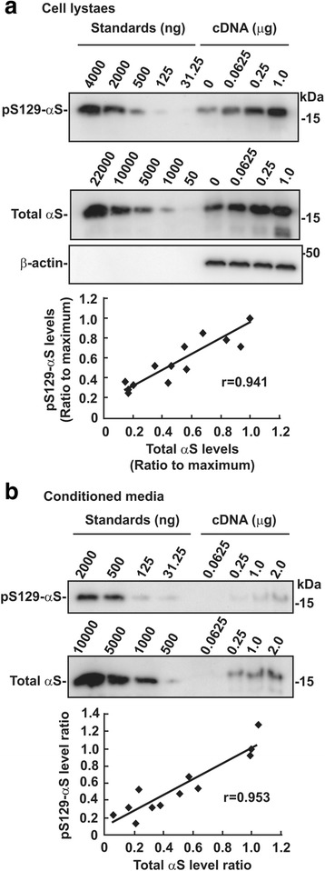
Parkinson's disease (PD) is characterized neuropathologically by intracellular aggregates of fibrillar α-synuclein, termed Lewy bodies (LBs). Approximately 90% of α-synuclein deposited as LBs is phosphorylated at Ser129 in brains with PD. In contrast, only 4% of total α-synuclein is phosphorylated at Ser129 in brains with normal individuals. It is unclear why extensive phosphorylation occurs in the pathological process of PD. To address this issue, we investigated a mechanism and role of Ser129-phosphorylation in regulating accumulation of α-synuclein. In CHO cells, the levels of Ser129-phosphorylated soluble α-synuclein were maintained constantly to those of total α-synuclein in intracellular and extracellular spaces. In SH-SY5Y cells and rat primary cortical neurons, mitochondrial impairment by rotenone or MPP+ enhanced Ser129-phosphorylation through increased influx of extracellular Ca2+. This elevation was suppressively controlled by targeting Ser129-phosphorylated α-synuclein to the proteasome pathway. Rotenone-induced insoluble α-synuclein was also targeted by Ser129-phosphoryation to the proteasome pathway. Experiments with epoxomicin and chloroquine showed that proteasomal targeting of insoluble Ser129-phosphorylated α-synuclein was enhanced under lysosome inhibition and it reduced accumulation of insoluble total α-synuclein. However, in a rat AAV-mediated α-synuclein overexpression model, there was no difference in the number of total α-synuclein aggregates between A53T mutant and A53T plus S129A double mutant α-synuclein, although Ser129-phosphorylated α-synuclein-positive aggregates were increased in rats expressing A53T α-synuclein. These findings suggest that Ser129-phosphorylation occurs against stress conditions, which increases influx of extracellular Ca2+, and it prevents accumulation of insoluble α-synuclein by evoking proteasomal clearance complementary to lysosomal one. However, Ser129-phosphorylation may provide an ineffective signal for degradation-resistant aggregates, causing extensive phosphorylation in aggregates.

Keywords: Aggregation; Mitochondrial impairment; Parkinson’s disease; Phosphorylation; Proteasome pathway; α–Synuclein.


Figures

Fig. 1
Fig. 1
Relation of Ser129-phosphorylated α-syn levels to total α-syn ones in intra- and extracellular spaces. CHO cells were transfected with the empty vector or the indicated amounts of wild-type α-syn cDNA. Samples were loaded along with recombinant α-syn proteins and Ser129-phosphorylated α-syn proteins for standards, followed by western blotting. Bands of Ser129-phosphorylated α-syn and total α-syn, including phosphorylated and non-phosphorylated forms, were detected by EP1536Y and Syn-1 antibody, respectively. Relative band intensities of Ser129-phosphorylated α-syn and total α-syn were corrected by plotting them on the standard curves, and then normalized to the intensities of β-actin. a Relation in intracellular α-syn. Cell lystaes (10 μg/lane) were loaded. b Relation in extracellular α-syn. After TCA-precipitated proteins were resolved by Laemmli’s sample buffer, samples corresponding to 20% of CM volume were loaded. Graphs show the positive correlation between Ser-129 phosphorylated and total α-syn levels
Fig. 2
Fig. 2
Effects of Ca2+ on Ser129-phosphorylation of α-syn. SH-SY5Y cell lines stably expressing wild-type α-syn (wt-aS/SH #4) were incubated in media containing 5 μM calcium ionophore A23187 except b. As vehicle control, cells were treated with DMSO at the same final concentration as reagents used. Cell lysates (15 μg/lane) were loaded on SDS-PAGE and analyzed by western botting with EP1536Y, Syn-1, or anti-β-actin (AC-15) antibody. a Effect of A23187 incubation time on Ser129-phosphorylation. Cells were treated with A23187 for the indicated time points until 8 h. b Effect of A23187 concentrations on Ser129-phosphorylation. Cells were treated with A23187 at the indicated concentrations for 8 h. c, d Effect of extracellular Ca2+ chelator EGTA (c) or intracellular Ca2+ chelator BAPTA-AM (B-AM) (d) on A23187-induced Ser129-phosphorylation. Cells were incubated in media containing 5 μM A23187 with the indicated concentrations of EGTA or BAPTA-AM for 4 h. e, f Effect of CaM inhibitor W-7 (e) or calmidazolium (Calm) (f) on A23187-induced Ser129-phosphorylation. Cells were incubated in media containing A23187 with the indicated concentrations of W-7 or calmidazolium for 4 h. Representative blots are shown. In the graphs of a to f, relative band intensities of Ser129-phosphorylated α-syn and total α-syn were normalized to those of β-actin. Data represent means ± SD and P values were estimated by one-way ANOVA with Bonferroni correction or Welch-ANOVA with Games-Howell post hoc test for unequal-variances (*, P < 0.05; **, P < 0.01)
Fig. 3
Fig. 3
Effects of mitochondrial complex I inhibitors MPP+ and rotenone on Ser129-phosphorylation of α-syn. Cell lysates (10 μg/lane) were loaded on SDS-PAGE and analyzed by western botting with EP1536Y, Syn-1, or AC-15 antibody. a Effect of incubation time of MPP+ and rotenone on Ser129-phosphorylation. Wt-aS/SH #4 cells were incubated in media containing 1 mM MPP+ or 5 μM rotenone for the indicated time points until 16 h. Vehicle controls were treated with DMSO at the same final concentration as each reagent. b Effect of concentrations of MPP+ or rotenone on Ser129-phosphorylation. Cells were incubated in media containing the indicated amounts of MPP+ or rotenone for 12 h. Graphs show relative band intensities of Ser129-phosphorylated α-syn and total α-syn. They were normalized to those of β-actin. Data represent means ± SD and P values were estimated by one-way ANOVA with Bonferroni correction or Welch-ANOVA with Games-Howell post hoc test for unequal-variances (*, P < 0.05; **, P < 0.01)
Fig. 4
Fig. 4
Role of Ca2+ and CaM in mitochondria complex I inhibitor-induced Ser129-phosphorylation of α-syn. As vehicle control, cells were treated with DMSO at the same final concentration as reagents used. Cell lysates (10 μg/lane) were loaded on SDS-PAGE and analyzed by western botting with EP1536Y, Syn-1, or AC-15 antibody. a, b Effect of intracellular Ca2+ chelator BAPTA-AM (a) or extracelluar Ca2+ chelator EGTA (b) on MPP+-induced Ser129-phosphorylation. Wt-aS/SH #4 cells were incubated in media containing 1 mM MPP+ and the indicated concentrations of BAPTA-AM or EGTA for 16 h. c, d Effect of BAPTA-AM (c) and EGTA (d) on rotenone-induced Ser129-phosphorylation. Wt-aS/SH #4 cells were incubated in media containing 5 μM rotenone and the indicated concentrations of BAPTA-AM or EGTA for 16 h. e, f Effect of CaM inhibitor W-7 on MPP+ (e)- or rotenone (f)-induced Ser129-phosphorylation. Cells were incubated in media containing 1 mM MPP+ or 5 μM rotenone with W-7 at the indicated concentrations for 16 h. Representative blots are shown. In the graphs of a to f, relative band intensities of Ser129-phosphorylated α-syn and total α-syn were normalized to those of β-actin. Data represent means ± SD and P values were estimated by one-way ANOVA with Bonferroni correction (*, P < 0.05; **, P < 0.01)
Fig. 5
Fig. 5
Ser129-mediated proteasomal targeting of soluble α-syn in mitochondrial complex I inhibition by rotenone. In each treatment, the concentration of DMSO was prepared to be equal. Cell lysates (2.5 μg/lane) were loaded on SDS-PAGE and analyzed by western botting with EP1536Y, Syn-1, or AC-15 antibody. a CHX-chase experiments for analyzing alteration in the metabolic fates of Ser129-phosphorylated and total α-syn by MG132 treatment. Wt-aS/SH #4 cells were pre-incubated in media containing either DMSO or 10 μM rotenone for 8 h. Then, they were incubated in fresh media further containing 100 μM cycloheximide (CHX) and/or 10 μM MG132 until 120 min. b Comparison of metabolic fates of rotenone-induced Ser129-phosphorylated α-syn with physiologically phosphorylated α-syn. Cells were pre-incubated in media containing DMSO or 10 μM rotenone for 8 h before CHX-chase experiments. Representative blots are shown. In a and b, graphs show the metabolic fates of Ser129-phosphorylated α-syn (left) and total α-syn (right). Black, yellow, blue and red lines represent treatment with DMSO, rotenone, MG132 and rotenone plus MG132, respectively. Data show means ± SD and P values were estimated by one-way ANOVA with Bonferroni correction (*, P < 0.05; **, P < 0.01)
Fig. 6
Fig. 6
Effect of Ser129-phosphorylation on α-syn solubility change by mitochondrial complex I inhibition. Wt-aS/SH #4 cells were fractionated into 1% Triton X-100 soluble and insoluble fractions by centrifugation at 100,000×g for 30 min. 1% Triton X-100 insoluble pellets were resolved by 8 M urea / 2% SDS solution. In 1% Triton X-100 soluble fractions, the extract (2.5 μg / lane) were loaded onto SDS-PAGE. In 1% Triton X-100 insoluble fractions, samples corresponding to 15 μg of soluble fractions were loaded. These samples were analyzed by western botting with EP1536Y, Syn-1, or AC-15 antibody. a Solubility change of α-syn by rotenone treatment. Cells were incubated by 10 or 50 nM rotenone for 5 days. The representative blots are shown. b CHX-chase experiments for analyzing alteration in the metabolic fates of insoluble Ser129-phosphorylated and total α-syn by MG132 treatment. After treatment with 50 nM rotenone for 5 days, cells were incubated in fresh media further containing 100 μM cycloheximide (CHX) and either 0.1% DMSO or 10 μM MG132 until 120 min. Graphs show the metabolic fates of Ser129-phosphorylated α-syn (left) and total α-syn (right). Data show means ± SD and P values were estimated by one-way ANOVA with Bonferroni correction (*, P < 0.05; **, P < 0.01)
Fig. 7
Fig. 7
Relation of Ser129-phosphorylation-mediated α-syn clearance between the proteasome and lysosome pathways. Wt-aS/SH #4 cells were fractionated into 1% Triton X-100 soluble and insoluble fractions by centrifugation at 100,000×g for 30 min. In 1% Triton X-100 soluble fractions, the extract (2.5 μg / lane) were loaded onto SDS-PAGE. In 1% Triton X-100 insoluble fractions, samples corresponding to 15 μg of soluble fractions were loaded. Samples were analyzed by western botting with EP1536Y, Syn-1, or AC-15 antibody. a Effect of proteasomal and lysosomal inhibitions on the metabolism of α-syn. Cells were incubated by either 100 nM epoxomicin, 100 μM chloroquine, or 100 nM epoxomicin plus 100 μM chloroquine for 24 h. Vehicle control cells were treated with the same concentration of DMSO. Upper and lower panels show the blots of 1% Triton X-100 insoluble fractions and 1% Triton X-100 soluble fractions, respectively. b Effect of Ser129-phosphorylation on the metabolism of α-syn in proteasomal and lysosomal inhibitions. Wt-aS/SH #4 cells and S129A-aS/SH #10 cells were incubated by either 100 nM epoxomicin, 100 μM chloroquine, or epoxomicin plus chloroquine for 24 h. Upper panels show the blots of 1% Triton X-100 insoluble fractions. Graph shows relative ratios of total α-syn to vehicle control cells. Data represent means ± SD and P values were estimated by one-way ANOVA with Bonferroni correction (*, P < 0.05; **, P < 0.01)
Fig. 8
Fig. 8
Effect of Ser129-phosphorylation on α-syn aggregate formation in a rat AAV-mediated α-syn overexpression model. Rats were sterotaxically injected with rAAV particles into the substantia nigra, and they expressed A53T mutant α-syn or A53T plus S129A double mutant α-syn. These rats were used in our previous study [18]. Information on the expression levels and toxicity is described in this paper [18]. Upper panels show the photomicrographs of rat striatum immunohistochemically stained with antibody specific to Ser129-phosphorylated α-syn and human total α-syn (LB509). We counted the number of α-syn-positive aggregates larger than 5 μm in diameter. Arrow heads indicate α-syn aggregates. Bar shows 100 μm. Graph shows quantitative analysis of striatal aggregates containing Ser129-phosphorylated α-syn or total α-syn. Data represent means ± SD and P values were estimated by one-way ANOVA with Bonferroni correction (*, P < 0.05; **, P < 0.01)
Fig. 9
Fig. 9
A model of Ser129-phosphorylation role in regulating α-syn levels and forming α-syn aggregates. Mitochondrial impairment stimulates solubility change of α-syn proteins from normally soluble forms to insoluble forms. Also, mitochondrial impairment facilitates Ser129-phosphorylation of α-syn by an increase in influx of extracellular Ca2+. Ser129-phosphorylated α-syn, including soluble and insoluble forms, is targeted to the proteasome pathway. Proteasomal targeting of Ser129-phosphorylated α-syn is more promoted under lysosome inhibition. It acts as a suppressor complementary to the lysosome pathway against accumulation of insoluble α-syn proteins. Also, α-syn aggregates undergo Ser129-phosphorylation. However, Ser129-phosphorylation-mediated proteasomal targeting is ineffective, once α-syn aggregates turn to be degradation-resistant. Consequently, α-syn proteins deposited in aggregates are extensively phosphorylated

References

    1. Anderson JP, Walker DE, Goldstein JM, de Laat R, Banducci K, Caccavello RJ, Barbour R, Huang J, Kling K, Lee M, et al. Phosphorylation of ser-129 is the dominant pathological modification of alpha-synuclein in familial and sporadic Lewy body disease. J Biol Chem. 2006;281:29739–29752. doi: 10.1074/jbc.M600933200. - DOI - PubMed
    1. Arawaka S, Fukushima S, Sato H, Sasaki A, Koga K, Koyama S, Kato T (2014) Zonisamide attenuates alpha-synuclein neurotoxicity by an aggregation-independent mechanism in a rat model of familial Parkinson’s disease. PLoS One 9:e89076. https://doi.org/10.1371/journal.pone.0089076 - DOI - PMC - PubMed
    1. Bouchard MJ, Wang LH, Schneider RJ. Calcium signaling by HBx protein in hepatitis B virus DNA replication. Science. 2001;294:2376–2378. doi: 10.1126/science.294.5550.2376. - DOI - PubMed
    1. Chen L, Feany MB. Alpha-synuclein phosphorylation controls neurotoxicity and inclusion formation in a Drosophila model of Parkinson disease. Nat Neurosci. 2005;8:657–663. doi: 10.1038/nn1443. - DOI - PubMed
    1. Cuervo AM, Stefanis L, Fredenburg R, Lansbury PT, Sulzer D. Impaired degradation of mutant alpha-synuclein by chaperone-mediated autophagy. Science. 2004;305:1292–1295. doi: 10.1126/science.1101738. - DOI - PubMed

Publication types

MeSH terms

LinkOut - more resources