Review
. 2017 Oct:139:123-137.
doi: 10.1016/j.visres.2017.05.009. Epub 2017 Jun 27.

The inner blood-retinal barrier: Cellular basis and development

Affiliations
Review

The inner blood-retinal barrier: Cellular basis and development

Mónica Díaz-Coránguez et al. Vision Res. 2017 Oct.

Abstract

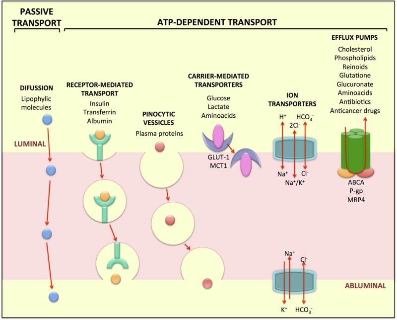
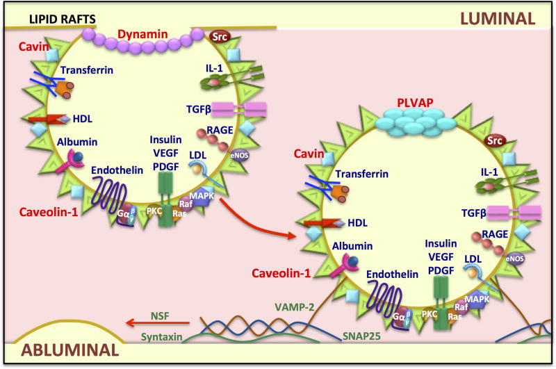
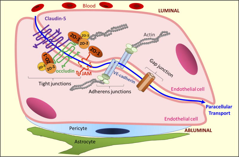
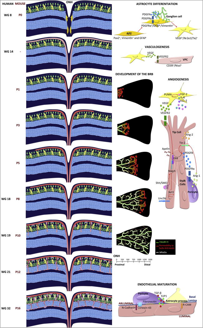
The blood-retinal barrier (BRB) regulates transport across retinal capillaries maintaining proper neural homeostasis and protecting the neural tissue from potential blood borne toxicity. Loss of the BRB contributes to the pathophysiology of a number of blinding retinal diseases including diabetic retinopathy. In this review, we address the basis of the BRB, including the molecular mechanisms that regulate flux across the retinal vascular bed. The routes of transcellular and paracellular flux are described as well as alterations in these pathways in response to permeabilizing agents in diabetes. Finally, we provide information on exciting new studies that help to elucidate the process of BRB development or barriergenesis and how understanding this process may lead to new opportunities for barrier restoration in diabetic retinopathy.

Keywords: Barriergenesis; Blood-retinal barrier; Paracellular permeability; Tight junctions; Transcytosis.


Figures

Figure 1
Figure 1. Mechanisms of transcellular transport across retinal endothelial cells
Some molecules can cross by diffusion due to their lipophilic properties. Other transport mechanisms are energy-dependent processes and include receptor-mediated transport, pinocytic vesicles, carrier-mediated transporters, ion transporters and efflux pumps. The endothelial cells that constitute the BRB, express a low number of these transporter mechanisms, some of the most important are indicated.
Figure 2
Figure 2. Molecular mechanisms in caveolae-mediated transport
Caveolae are lipid rafts or membrane microdomains enriched in cholesterol that promote the clustering of molecules at the membrane. Caveolin-1 is enriched in these domains and caveolae are responsible for vesicle dependent transport in endothelial cells. Other molecules involved in caveolae formation and transcytosis are indicated in red. Caveolae vesicles also contain receptors (blue) and signaling molecules (white) that can transduce an external signal to the cell interior. Caveolae are able to recycle to the luminal membrane or to regulate transport to the abluminal membrane. Finally, vesicle fusion proteins (green) including the SNAREs VAMP-2 and SNAP25, regulate fusion to the membrane.
Figure 3
Figure 3. Paracellular Transport
In the BRB, molecules can move from the blood (luminal) into the retinal (abluminal) side via the paracellular space, which is located in between two adjacent cells (blue line). Some of the molecules central to control of paracellular transport of the BRB are indicated.
Figure 4
Figure 4. Development of retinal vasculature and the inner blood-retinal barrier
Left panel, retinal cross sections demonstrating vascular development and indicated times, middle panel shows the development of the iBRB, and the right panel depicts the molecular mechanisms regulating angiogenesis or barriergenesis. During retinal vascular development, astrocyte precursor cells (APC in gold) emerge from the optic nerve head and migrate towards the PDGF secreted by ganglion cells (green). The astrocytes (yellow) differentiation program starts once the APCs leave the ONH and emerge into the retinal tissue. Inside the retina, the hypoxic environment induces astrocytes to secrete VEGF. In humans at WG14, the vascular patent cells (VPC; light pink) migrate into the retina expanding though the perifoveal region. VPCs differentiate to form the first vascular plexus layer. In rodents, this process has not been observed, instead vascular sprouting across the retinal ganglion layer is observed (P8). Astrocytes release additional chemotactic factors that promote endothelial cell sprouting into the deep layers of the retina (P10-P12). Finally, pericyte recruitment and glial processes interact with the vascular endothelium to create the neurovascular unit (WG32, P16). The development of the iBRB occurs at the same time from P1 to P10. The new sprouts show leakiness (red color in middle pannel) while the stalk cells located closer to the ONH have already developed a functional BRB. This barriergenesis correlates with Msfsd2a expression (white). All endothelial cells express claudin-5 (green).

References

    1. Alvarez JI, Dodelet-Devillers A, Kebir H, Ifergan I, Fabre PJ, Terouz S, Sabbagh M, Wosik K, Bourbonniere L, Bernard M, van Horssen J, de Vries HE, Charron F, Prat A. The Hedgehog pathway promotes blood-brain barrier integrity and CNS immune quiescence. Science. 2011;334(6063):1727–1731. - PubMed
    1. Anderson KD, Pan L, Yang XM, Hughes VC, Walls JR, Dominguez MG, Simmons MV, Burfeind P, Xue Y, Wei Y, Macdonald LE, Thurston G, Daly C, Lin HC, Economides AN, Valenzuela DM, Murphy AJ, Yancopoulos GD, Gale NW. Angiogenic sprouting into neural tissue requires Gpr124, an orphan G protein-coupled receptor. Proc Natl Acad Sci U S A. 2011;108(7):2807–2812. - PMC - PubMed
    1. Anderson RG. Caveolae: where incoming and outgoing messengers meet. Proc Natl Acad Sci U S A. 1993;90(23):10909–10913. - PMC - PubMed
    1. Andreone BJ, Chow BW, Tata A, Lacoste B, Ben-Zvi A, Bullock K, Deik AA, Ginty DD, Clish CB, Gu C. Blood-Brain Barrier Permeability Is Regulated by Lipid Transport-Dependent Suppression of Caveolae-Mediated Transcytosis. Neuron. 2017;94(3):581–594. e585. - PMC - PubMed
    1. Angelow S, Yu AS. Structure-function studies of claudin extracellular domains by cysteine-scanning mutagenesis. J Biol Chem. 2009;284(42):29205–29217. - PMC - PubMed

MeSH terms