. 2017 Sep 6;25(9):1999-2013.
doi: 10.1016/j.ymthe.2017.05.015. Epub 2017 Jun 12.

Using CRISPR-Cas9 to Generate Gene-Corrected Autologous iPSCs for the Treatment of Inherited Retinal Degeneration

Affiliations

Using CRISPR-Cas9 to Generate Gene-Corrected Autologous iPSCs for the Treatment of Inherited Retinal Degeneration

Erin R Burnight et al. Mol Ther. .

Abstract

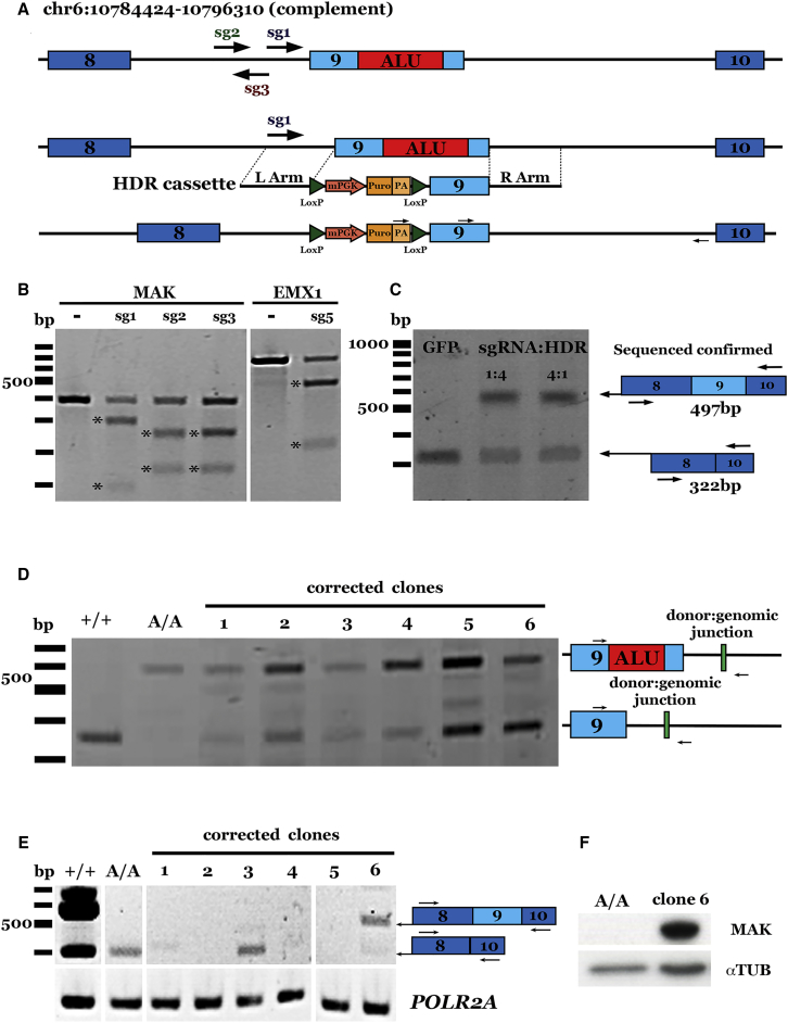
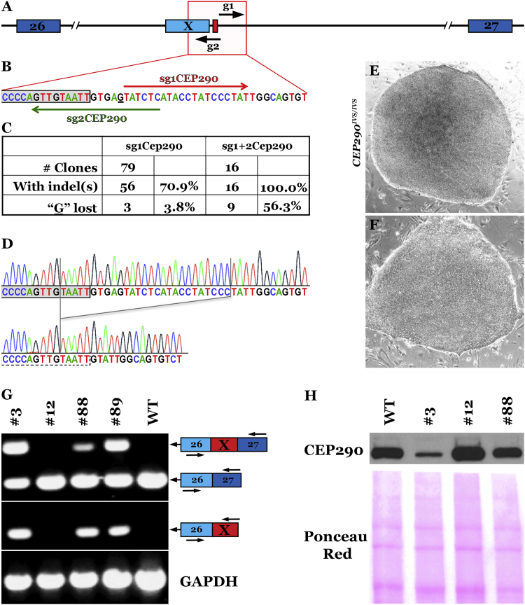
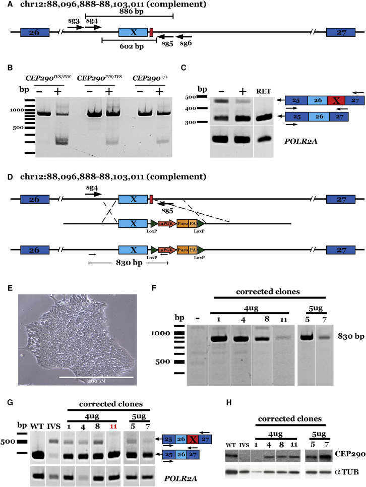
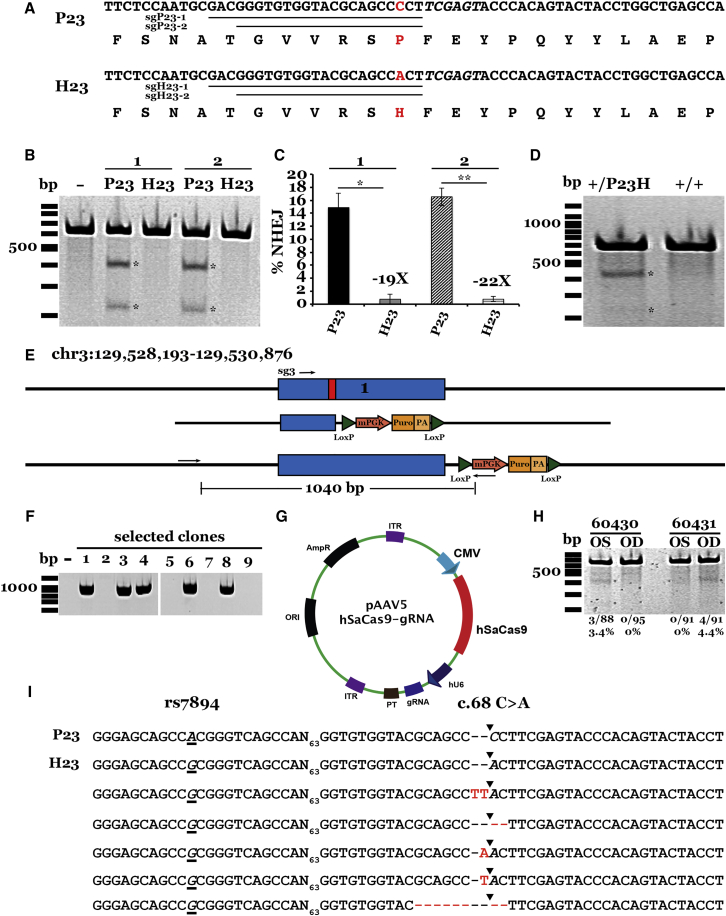
Patient-derived induced pluripotent stem cells (iPSCs) hold great promise for autologous cell replacement. However, for many inherited diseases, treatment will likely require genetic repair pre-transplantation. Genome editing technologies are useful for this application. The purpose of this study was to develop CRISPR-Cas9-mediated genome editing strategies to target and correct the three most common types of disease-causing variants in patient-derived iPSCs: (1) exonic, (2) deep intronic, and (3) dominant gain of function. We developed a homology-directed repair strategy targeting a homozygous Alu insertion in exon 9 of male germ cell-associated kinase (MAK) and demonstrated restoration of the retinal transcript and protein in patient cells. We generated a CRISPR-Cas9-mediated non-homologous end joining (NHEJ) approach to excise a major contributor to Leber congenital amaurosis, the IVS26 cryptic-splice mutation in CEP290, and demonstrated correction of the transcript and protein in patient iPSCs. Lastly, we designed allele-specific CRISPR guides that selectively target the mutant Pro23His rhodopsin (RHO) allele, which, following delivery to both patient iPSCs in vitro and pig retina in vivo, created a frameshift and premature stop that would prevent transcription of the disease-causing variant. The strategies developed in this study will prove useful for correcting a wide range of genetic variants in genes that cause inherited retinal degeneration.

Keywords: CRISPR; iPSCs; retinal degeneration.


Figures

Figure 1
Figure 1
CRISPR-Based Correction of an Alu Insertion in MAK (A) Schematic representation of our CRISPR-Cas9/HDR strategy for the correction of the Alu insertion (top; red box) in MAK. sgRNAs tested are indicated by arrows. A donor plasmid (middle; HDR cassette) containing 500 bp of homology flanking a floxed puromycin selection cassette and the wild-type exon 9 sequence was co-transfected using various ratios of sg1-SpCas9-expressing plasmid, resulting in the repaired allele (bottom). (B) Representative gel image of T7E1 assays in HEK293T cells for each MAK sgRNA developed; a previously reported sgRNA targeting the EMX1 locus was included as a control. (C) Representative gel image demonstrating restoration of wild-type MAK transcript in sg1-SpCas9-treated, puromycin-selected, iPSC-derived photoreceptor precursor cells from a patient with molecularly confirmed MAK-associated retinitis pigmentosa. (D) Representative gel image confirming HDR in selected iPSC clones. +/+ indicates unaffected control cells; A/A indicates uncorrected patient iPSCs. (E) Representative gel image demonstrating restoration of wild-type MAK transcript in one puromycin-selected iPSC clone (clone 6) from a patient with molecularly confirmed MAK-associated retinitis pigmentosa. (F) Representative gel image confirming restoration of MAK protein expression in corrected iPSC clone 6.
Figure 2
Figure 2
CRISPR-Cas9 Correction of the Deep Intronic IVS26 Mutation in CEP290 (A) Location of the two single guide RNAs. (B) sgRNA1 and 2 were designed to generate S. pyogenes Cas9-induced deletions that remove the mutant nucleotide and normalize CEP290 pre-mRNA splicing in hiPSCs homozygous for the CEP290 c.2991+1655A > G mutation (nucleotide 1655 of intron 26 is underlined). The gray box represents the 3′ end of the cryptic exon X. (C) Quantification of indel formation frequency and loss of the CEP290 disease-causing mutation with the single- or dual-guide strategy. (D–F) Representative sequence alignments (D) and phase-contrast images (4×) of two CEP290 clones: clone #3 (E), unchanged, and clone #12 (F), homozygous deletion. (G) CEP290 transcript analysis by semiquantitative RT-PCR. (Top) Amplicons obtained using primers located in exons 26 and 27 (E26, E27; EX represents inclusion of the cryptic exon X). (Bottom) Amplicons obtained with primers located in exons 26 and X. GAPDH was amplified as a control. (H) Western blot demonstrating the increase in full-length CEP290 protein expression in LCA-iPSCs with no (clone #3), one (clone #88), or two (clone #12) repaired alleles. Nitrocellulose Ponceau S. staining is shown to demonstrate even sample processing and loading.
Figure 3
Figure 3
CRISPR-Cas9 Correction of the Deep Intronic IVS26 Mutation in CEP290 Using S. aureus Cas9 (A) Schematic representation of our paired guide strategy for the correction of the deep intronic IVS26 cryptic splice site mutation in CEP290. Two guides targeting upstream (sg3 and sg4) of the IVS26 mutation (red box) were each paired with two guides (sg5 and sg6) targeting downstream of the mutation to delete the exon X sequence and IVS26 mutation. (B) Representative gel image showing sg4/5-SaCas9-mediated IVS26 deletion in patient-derived iPSCs. Two lines of patient-derived iPSCs carrying homozygous IVS26 genotypes and those from a control unaffected individual were transfected with sg4/5-SaCas9-expressing plasmid (+) or Cas9 only (−). (C) Representative gel image demonstrating restoration of CEP290 transcript in sg4/5-SaCas9-treated iPSCs. POLR2A was amplified as a loading control. RET, human control retina cDNA. (D) Schematic representation of our CRISPR-Cas9/HDR strategy for the correction of the IVS26 cryptic splice site mutation (top; red box) in CEP290. Sg4/5RNA pair-expressing plasmid was co-delivered with a donor plasmid (middle; HDR cassette) containing 500 bp of homology flanking a floxed puromycin selection cassette and the wild-type IVS26 sequence resulting in the repaired allele (bottom). (E) Bright field image of a homozygous IVS26 patient-derived iPSC clone post-puromycin selection (note normal feeder-free iPSC morphology). Scale bar, 400 μM. (F) Representative gel image of genomic DNA from iPSC clones co-transfected with sg4/5-expressing plasmid and donor plasmid and cultured under puromycin selection. (G) Representative gel image demonstrating restoration of CEP290 transcript in sg4/5-SaCas9/donor-plasmid-treated iPSC clones. POLR2A was amplified as a loading control. WT, unaffected control iPSC cDNA; IVS, uncorrected patient-derived iPSCs carrying homozygous IVS26 alleles. (H) Western blot demonstrating restoration of full-length CEP290 protein in CRISPR/HDR-treated iPSC clones. Anti-α-Tub was used as a loading control.
Figure 4
Figure 4
Allele-Specific Targeting of the Dominant Gain-of-Function Pro23His Mutation in RHO (A) Nucleotide and amino acid sequences of unaffected (P23) and affected (H23) RHO alleles. Underlined sequences indicate guides targeting each allele. Red text indicates mutation (c.68 C > A transversion at codon 23) lying in the seed region of each guide. (B) Representative gel image of T7E1 assays in HEK293T cells for each RHO sgRNA tested. (C) Histogram showing percent NHEJ in HEK293T cells transfected with each guide. Efficiency was quantified by subcloning and Sanger sequencing (please refer to Materials and Methods for further details). At lest 61 clones were sequenced per transfection. Data are represented as mean ± SEM. n = 3; *p < 0.01; **p < 0.001. (D) Representative gel image of T7E1 assays in patient-derived iPSCs (+/P23H) and iPSCs from an unaffected individual (+/+) transfected with plasmids expressing the sgH23-2 guide and SaCas9. (E) Schematic representation of our CRISPR-Cas9/HDR strategy for the correction of the Pro23His mutation (top; red box) in RHO. Sg3-hSpCas9-expressing plasmid was co-delivered with a donor plasmid (middle; HDR cassette) containing 500 bp of homology flanking a floxed puromycin selection cassette and the wild-type (Pro) sequence, resulting in the repaired allele (bottom). (F) Representative gel image of genomic DNA from iPSC clones co-transfected with sg3-expressing plasmid and donor plasmid and cultured under puromycin selection. (G) Schematic of transgene cassette plasmid carrying the allele-specific guide H23-2 and the humanized SaCas9 expression cassettes flanked by AAV2 ITRs packaged into the AAV5 serotype vector. (H) Representative gel image of T7E1 assays performed on genomic DNA isolated from the retinas of two animals treated unilaterally with AAV2/5-sgH23-2-SaCas9 vector. Amplicons were gel purified, subcloned, and sequenced via Sanger sequencing. Sequences from recovered clones (I) were aligned using Lasergene DNA analysis software. The percentage of alleles demonstrating NHEJ is indicated at the bottom of each lane. The rs7984 A/G SNP occurs 93 bp upstream of the Pro23His mutation. The P23 allele carries the rs7984 A nucleotide (top; underlined and italicized), whereas the H23 allele carries the rs7984 G nucleotide (second from top; underlined and italicized). Sequenced amplicon clones from sgH23-2-treated animals revealed formation of indel in the H23 allele only.

References

    1. Tucker B.A., Mullins R.F., Stone E.M. Stem cells for investigation and treatment of inherited retinal disease. Hum. Mol. Genet. 2014;23(R1):R9–R16. - PMC - PubMed
    1. Gregory-Evans K., Bhattacharya S.S. Genetic blindness: current concepts in the pathogenesis of human outer retinal dystrophies. Trends Genet. 1998;14:103–108. - PubMed
    1. Gilbert C., Foster A. Blindness in children: control priorities and research opportunities. Br. J. Ophthalmol. 2001;85:1025–1027. - PMC - PubMed
    1. Bush R.A., Hawks K.W., Sieving P.A. Preservation of inner retinal responses in the aged Royal College of Surgeons rat. Evidence against glutamate excitotoxicity in photoreceptor degeneration. Invest. Ophthalmol. Vis. Sci. 1995;36:2054–2062. - PubMed
    1. Santos A., Humayun M.S., de Juan E., Jr., Greenburg R.J., Marsh M.J., Klock I.B., Milam A.H. Preservation of the inner retina in retinitis pigmentosa. A morphometric analysis. Arch. Ophthalmol. 1997;115:511–515. - PubMed

MeSH terms

Substances