

IGFBP7 Deletion Promotes Hepatocellular Carcinoma
- PMID: 28619711
- PMCID: PMC5894280
- DOI: 10.1158/0008-5472.CAN-16-2885
IGFBP7 Deletion Promotes Hepatocellular Carcinoma
Abstract
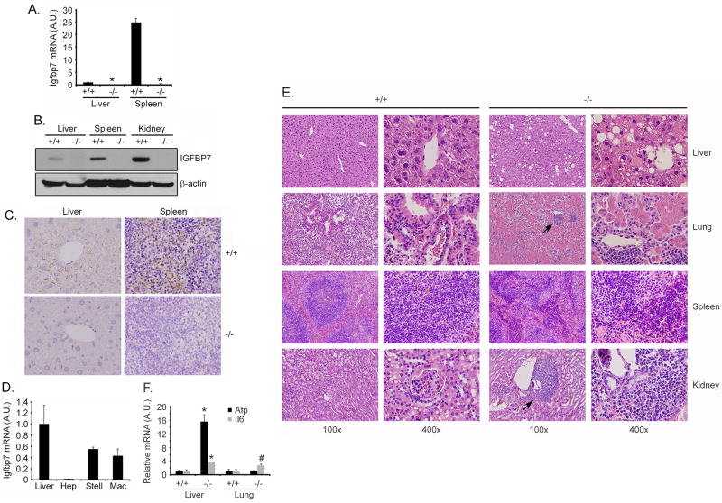
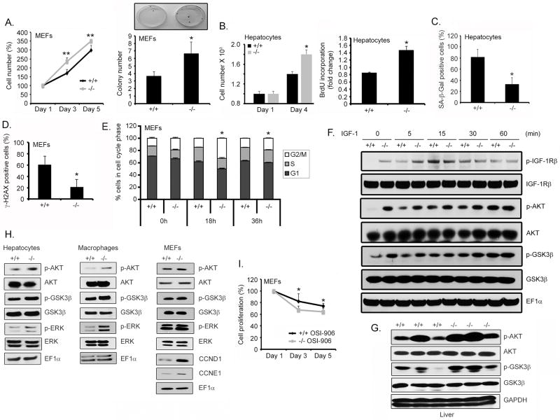
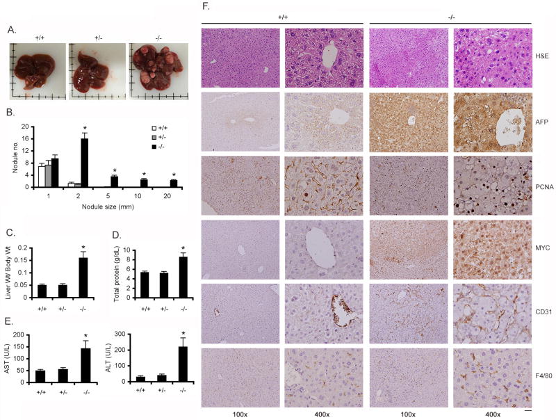
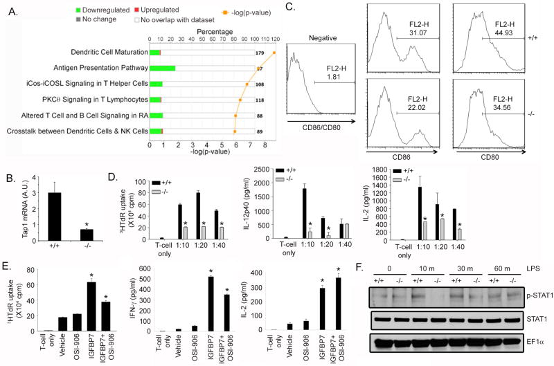
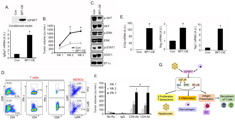
Activation of IGF signaling is a major oncogenic event in diverse cancers, including hepatocellular carcinoma (HCC). In this setting, the insulin-like growth factor binding protein IGFBP7 inhibits IGF signaling by binding the IGF1 receptor (IGF1R), functioning as a candidate tumor suppressor. IGFBP7 abrogates tumors by inhibiting angiogenesis and inducing cancer-specific senescence and apoptosis. Here, we report that Igfbp7-deficient mice exhibit constitutively active IGF signaling, presenting with proinflammatory and immunosuppressive microenvironments and spontaneous liver and lung tumors occurring with increased incidence in carcinogen-treated subjects. Igfbp7 deletion increased proliferation and decreased senescence of hepatocytes and mouse embryonic fibroblasts, effects that were blocked by treatment with IGF1 receptor inhibitor. Significant inhibition of genes regulating immune surveillance was observed in Igfbp7-/- murine livers, which was associated with a marked inhibition in antigen cross-presentation by Igfbp7-/- dendritic cells. Conversely, IGFBP7 overexpression inhibited growth of HCC cells in syngeneic immunocompetent mice. Depletion of CD4+ or CD8+ T lymphocytes abolished this growth inhibition, identifying it as an immune-mediated response. Our findings define an immune component of the pleiotropic mechanisms through which IGFBP7 suppresses HCC. Furthermore, they offer a genetically based preclinical proof of concept for IGFBP7 as a therapeutic target for immune management of HCC. Cancer Res; 77(15); 4014-25. ©2017 AACR.
©2017 American Association for Cancer Research.
Conflict of interest statement
Figures

Similar articles
-
Insulin-like growth factor-binding protein-7 functions as a potential tumor suppressor in hepatocellular carcinoma.Clin Cancer Res. 2011 Nov 1;17(21):6693-701. doi: 10.1158/1078-0432.CCR-10-2774. Epub 2011 Sep 9. Clin Cancer Res. 2011. PMID: 21908579 Free PMC article.
-
IGFBP7 downregulation is associated with tumor progression and clinical outcome in hepatocellular carcinoma.Int J Cancer. 2012 Jan 15;130(2):319-27. doi: 10.1002/ijc.25994. Epub 2011 Apr 13. Int J Cancer. 2012. PMID: 21328580
-
IGFBP7 binds to the IGF-1 receptor and blocks its activation by insulin-like growth factors.Sci Signal. 2012 Dec 18;5(255):ra92. doi: 10.1126/scisignal.2003184. Sci Signal. 2012. PMID: 23250396
-
Emerging role of insulin-like growth factor-binding protein 7 in hepatocellular carcinoma.J Hepatocell Carcinoma. 2014 Mar 26;1:9-19. doi: 10.2147/JHC.S44460. eCollection 2014. J Hepatocell Carcinoma. 2014. PMID: 27508172 Free PMC article. Review.
-
Insulin Growth Factor Binding Protein 7 (IGFBP7)-Related Cancer and IGFBP3 and IGFBP7 Crosstalk.Front Oncol. 2020 May 15;10:727. doi: 10.3389/fonc.2020.00727. eCollection 2020. Front Oncol. 2020. PMID: 32500027 Free PMC article. Review.
Cited by
-
A strategy for the treatment of gastrointestinal cancer: Targeting tumor senescent cells.Front Mol Biosci. 2023 Mar 6;10:1139840. doi: 10.3389/fmolb.2023.1139840. eCollection 2023. Front Mol Biosci. 2023. PMID: 36950520 Free PMC article. Review.
-
Comprehensive Analysis of IGFBPs as Biomarkers in Gastric Cancer.Front Oncol. 2021 Oct 21;11:723131. doi: 10.3389/fonc.2021.723131. eCollection 2021. Front Oncol. 2021. PMID: 34745945 Free PMC article.
-
Identification of a long noncoding RNA signature to predict outcomes of glioblastoma.Mol Med Rep. 2019 Jun;19(6):5406-5416. doi: 10.3892/mmr.2019.10184. Epub 2019 Apr 24. Mol Med Rep. 2019. PMID: 31059035 Free PMC article.
-
Expression characteristics and their functional role of IGFBP gene family in pan-cancer.BMC Cancer. 2023 Apr 24;23(1):371. doi: 10.1186/s12885-023-10832-3. BMC Cancer. 2023. PMID: 37088808 Free PMC article.
-
Bioinformatic identification and analysis of immune-related chromatin regulatory genes as potential biomarkers in idiopathic pulmonary fibrosis.Ann Transl Med. 2022 Aug;10(16):896. doi: 10.21037/atm-22-3700. Ann Transl Med. 2022. PMID: 36111015 Free PMC article.
References
-
- Neefjes J, Jongsma ML, Paul P, Bakke O. Towards a systems understanding of MHC class I and MHC class II antigen presentation. Nat Rev Immunol. 2011;11:823–36. - PubMed
-
- Makarova-Rusher OV, Medina-Echeverz J, Duffy AG, Greten TF. The yin and yang of evasion and immune activation in HCC. J Hepatol. 2015;62:1420–9. - PubMed
-
- Scharf JG, Braulke T. The role of the IGF axis in hepatocarcinogenesis. Hormone Metab Res. 2003;35:685–93. - PubMed
Publication types
MeSH terms
Substances
Grants and funding
LinkOut - more resources
Full Text Sources
Other Literature Sources
Medical
Molecular Biology Databases
Research Materials
Miscellaneous
