. 2017 Jul 15;144(14):2652-2662.
doi: 10.1242/dev.147751. Epub 2017 Jun 15.

Convergence of signaling pathways underlying habenular formation and axonal outgrowth in zebrafish

Affiliations

Convergence of signaling pathways underlying habenular formation and axonal outgrowth in zebrafish

Sara Roberson et al. Development. .

Abstract

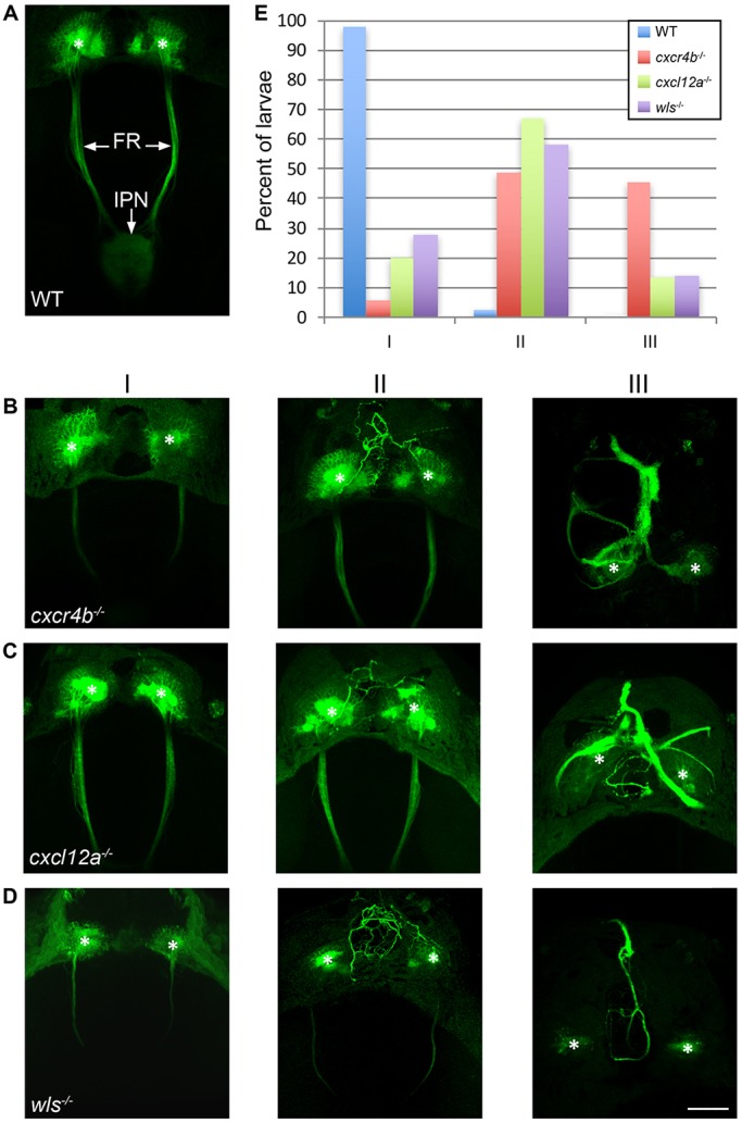
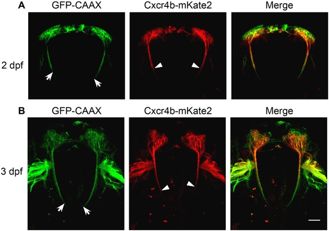
The habenular nuclei are a conserved integrating center in the vertebrate epithalamus, where they modulate diverse behaviors. Despite their importance, our understanding of habenular development is incomplete. Time-lapse imaging and fate mapping demonstrate that the dorsal habenulae (dHb) of zebrafish are derived from dbx1b-expressing (dbx1b+ ) progenitors, which transition into cxcr4b-expressing neuronal precursors. The precursors give rise to differentiated neurons, the axons of which innervate the midbrain interpeduncular nucleus (IPN). Formation of the dbx1b+ progenitor population relies on the activity of the Shh, Wnt and Fgf signaling pathways. Wnt and Fgf function additively to generate dHb progenitors. Surprisingly, Wnt signaling also negatively regulates fgf8a, confining expression to a discrete dorsal diencephalic domain. Moreover, the Wnt and Fgf pathways have opposing roles in transcriptional regulation of components of the Cxcr4-chemokine signaling pathway. The chemokine pathway, in turn, directs the posterior outgrowth of dHb efferents toward the IPN and, when disrupted, results in ectopic, anteriorly directed axonal projections. The results define a signaling network underlying the generation of dHb neurons and connectivity with their midbrain target.

Keywords: Chemokine; Fgf; Habenula; Interpeduncular nucleus; Shh; Wnt.


Conflict of interest statement

Competing interestsThe authors declare no competing or financial interests.

Figures

Fig. 1.
Fig. 1.
Live imaging of dbx1b+ and cxcr4b+ populations. Live imaging of WT and wls mutant TgBAC(dbx1b:GFP) and TgBAC(cxcr4b:nls-dTomato) double-labeled embryos between 27 and 48 hpf (Movie 1). Black and white images are of dTomato-labeled nuclei alone. dTomato-labeled nuclei are found in the pineal anlage (dashed circle) and, over time, in cells arising within the developing dHb (arrowheads and inset). Scale bar: 30 µm (11 µm for inset).
Fig. 2.
Fig. 2.
Temporal switch in dbx1b and cxcr4b expression in the developing dHb. Relative positions of dbx1b+ and cxcr4b+ cell populations revealed by TgBAC(dbx1b:GFP) and TgBAC(cxcr4b:nls-dTomato) labeling, respectively, between 2 and 30 dpf. Merged images are shown in dorsal and frontal views. Scale bars: 30 µm.
Fig. 3.
Fig. 3.
Ventromedial position of cxcr4b-expressing cells relative to dHb neurons. Fluorescence in situ hybridization in 2 and 3 dpf larvae for transcripts of cxcr4b and (A) the pan-neuronal gene elavl3 or (B) ano2, a marker of dHb neurons. The left three columns of images are corresponding dorsal views; the rightmost column shows frontal views. Scale bar: 30 µm.
Fig. 4.
Fig. 4.
Specification of dbx1b+ dHb progenitors requires Shh signaling upstream of or parallel to Wnt signaling. At 36 hpf, (A,A′) cxcr4b and (B-C′) dbx1b transcripts are found in the developing dHb of (A,B,C) WT embryos, but not in the corresponding diencephalic region of (A′,B′,C′) smo mutants (arrowheads). Expression of (D-E′) shha and (F-G′) ptch1 is comparable in (D,E,F,G) WT embryos and (D′,E′,F′,G′) wls mutants. Sibling WT and homozygous wls mutant embryos are distinguished by the presence or absence of wls transcripts (Kuan et al., 2015). Dorsal (A-B′,D,D′,F,F′) and lateral (C,C′,E,E′,G,G′) views are shown. Scale bars: 30 µm.
Fig. 5.
Fig. 5.
Wnt signaling defines the domain of Fgf signaling in the dorsal diencephalon. (A-C′) At 35 hpf, the domain of fgf8a expression just rostral to the developing dHb is expanded in wls mutants. (D,D′) Inhibition of Wnt signaling at 36 hpf by heat shock activation of Tg(hsp70:dkk1-GFP) also expands fgf8a expression at 48 hpf. (E-H′) The expression domains of (E,F) dusp6 and (G,H) etv5b, two targets of Fgf signaling, are enlarged in (E′,F′,G′,H′) wls mutants (compare brackets). (I-L) Expression of dbx1b (arrowheads) in the (I) WT dorsal diencephalon is reduced in (J) fgf8a and (K) wls mutants, and absent in (L) fgf8a;wls double mutants. Dorsal (A,C,D,E,G) and lateral (B,F,H,I-L) views are shown. Refer to Table 1 for numerical values. Sibling WT and homozygous wls mutant embryos were distinguished by the presence or absence of wls transcripts (Kuan et al., 2015). Scale bars: 50 µm.
Fig. 6.
Fig. 6.
Fgf signaling regulates expression domains of chemokine pathway components. (A-F) The (A-C) cxcl12a and (D-F) cxcl12b genes are transcribed in regions adjacent to the dHb at 35 hpf, as shown by fluorescence in situ hybridization and immunolabeling for GFP in TgBAC(dbx1b:GFP) embryos. (G-H′) cxcl12b and (I-J′) ackr3b expression domains (brackets) are expanded in wls mutants. In fgf8a mutants, (K-L′) cxcl12b and (M-N′) ackr3b dorsal diencephalic expression domains (arrowheads) are reduced. Although the cxcl12b expression domain is reduced in (P) fgf8a mutants and expanded in (Q) wls mutants, (R) fgf8a;wls double mutants show a pattern more similar to (O) WT. Between 24 and 48 hpf, WT and wls mutant siblings were treated with either (S,S′) 0.3% DMSO, (T,T′) 18 µM SU5402+0.3% DMSO, or (U,U′) 24 µM SU5402+0.3% DMSO. Following treatment with 24 µM SU5402, cxcl12b expression in wls mutants is restored to the WT pattern (brackets). Scale bar: 30 µm. Refer to Table 1 for numerical values. Dorsal (A-G′,I,I′,K,K′,M,M′,S-U′) and lateral (H,H′,J,J′,L,L′,N,N′,O-R) views are shown. Scale bars: 50 µm.
Fig. 7.
Fig. 7.
Chemokine signaling directs outgrowth of dHb axons. (A) In 5 dpf WT larvae, TgBAC(gng8:CAAX-GFP) labels neurons in the left and right dHb and their efferent axons in the FR, which terminate at the midbrain IPN. (B-D) Axonal morphology of 5 dpf (B) cxcr4b, (C) cxcl12a and (D) wls mutants falls into three general phenotypic classes: in Class I, the majority of axons fasciculate normally and project posteriorly; in Class II, some ectopic axons extend anterior to the dHb; and in Class III, the majority of axons grow anteriorly and fasciculate as a thick midline bundle. Asterisks indicate the dHb. Scale bar: 50 µm. (E) Percentage of WT siblings (blue, n=41) and cxcr4b (red, n=35), cxcl12a (green, n=30) and wls (purple, n=43) mutants with Class I, II or III axonal morphology.
Fig. 8.
Fig. 8.
Chemokine signaling reporter labels newly born neurons and emerging axons. TgBAC(cxcr4b:cxcr4b-mKate2-IRES-GFP-CAAX) labeling at (A) 2 and (B) 3 dpf. GFP labels the membranes of dHb neurons and axons. The Cxcr4b chemokine receptor fusion protein (red) is found in neurons near the ventricular zone and on proximal (arrowheads) but not distal (arrows) regions of their axons. Scale bar: 50 µm.
Fig. 9.
Fig. 9.
Schematic of signaling pathways in dHb development. (A) Timeline indicating when dbx1b+ progenitors, cxcr4b+ precursors and ano2+ neurons are detected in the developing dHb. (B) In zebrafish embryos, Wnt and Fgf signaling function additively to generate dbx1b+ progenitors (purple), which produce cxcr4b+ neural precursors (yellow) that differentiate into habenular neurons (green). Shh acts upstream of or parallel to Wnt and Fgf signaling. The Wnt pathway also regulates the spatial extent of Fgf signaling, which in turn confines cxcl12b (light blue) and ackr3b (dark blue) transcription to regions anterior of the developing dHb. cxcl12a (gray) transcripts are present in bilateral domains posterior and ventral to the habenular region. Establishing this spatial pattern of chemokine signaling later influences the outgrowth of dHb efferents. (B′) In WT larvae, anterior inhibition or posterior attraction directs outgrowth of dHb axons caudally towards the IPN. (C) In wls mutants, Wnt signaling is disrupted, dbx1b+ progenitors are reduced, and the dHb are significantly smaller (Kuan et al., 2015). Additionally, Fgf signaling is expanded, as are cxcl12b and ackr3b domains of expression, resulting in ectopic chemokine signaling and (C′) aberrant rostral projections from dHb neurons. (D) In mutants lacking either the Cxcr4b chemokine receptor (dashed line) or the Cxcl12a chemokine (hatched area) formation of the dHb appears normal, but (D′) dHb axon outgrowth is aberrant.

References

    1. Aizawa H., Goto M., Sato T. and Okamoto H. (2007). Temporally regulated asymmetric neurogenesis causes left-right differences in the zebrafish habenular structures. Dev. Cell 12, 87-98. 10.1016/j.devcel.2006.10.004 - DOI - PubMed
    1. Aizawa H., Amo R. and Okamoto H. (2011). Phylogeny and ontogeny of the habenular structure. Front. Neurosci. 5, 138 10.3389/fnins.2011.00138 - DOI - PMC - PubMed
    1. Aman A. and Piotrowski T. (2008). Wnt/β-catenin and Fgf signaling control collective cell migration by restricting chemokine receptor expression. Dev. Cell 15, 749-761. 10.1016/j.devcel.2008.10.002 - DOI - PubMed
    1. Antolin-Fontes B., Ables J. L., Görlich A. and Ibañez-Tallon I. (2015). The habenulo-interpeduncular pathway in nicotine aversion and withdrawal. Neuropharmacology 96, 213-222. 10.1016/j.neuropharm.2014.11.019 - DOI - PMC - PubMed
    1. Baldwin P. R., Alanis R. and Salas R. (2011). The role of the habenula in nicotine addiction. J. Addict. Res. Ther. S1, 002 10.4172/2155-6105.S1-002 - DOI - PMC - PubMed

MeSH terms