. 2017 Jul 15;144(14):2673-2682.
doi: 10.1242/dev.148767. Epub 2017 Jun 15.

COP9 signalosome subunits protect Capicua from MAPK-dependent and -independent mechanisms of degradation

Affiliations

COP9 signalosome subunits protect Capicua from MAPK-dependent and -independent mechanisms of degradation

Annabelle Suisse et al. Development. .

Abstract

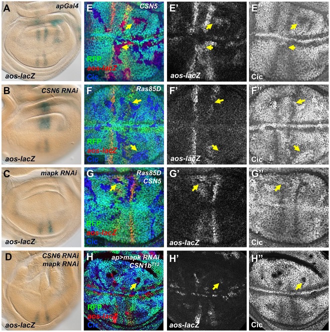
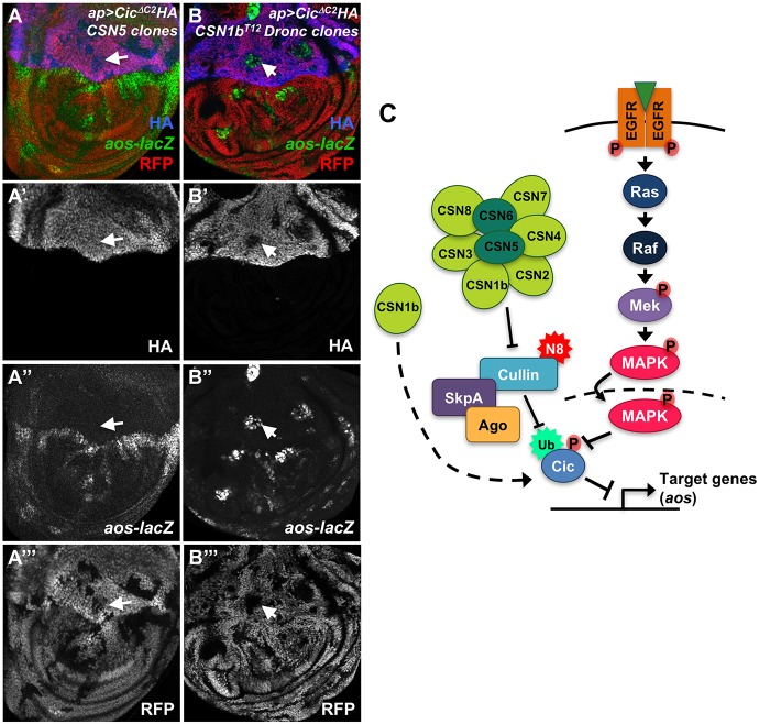
The COP9 signalosome removes Nedd8 modifications from the Cullin subunits of ubiquitin ligase complexes, reducing their activity. Here, we show that mutations in the Drosophila COP9 signalosome subunit 1b (CSN1b) gene increase the activity of ubiquitin ligases that contain Cullin 1. Analysis of CSN1b mutant phenotypes revealed a requirement for the COP9 signalosome to prevent ectopic expression of Epidermal growth factor receptor (EGFR) target genes. It does so by protecting Capicua, a transcriptional repressor of EGFR target genes, from EGFR pathway-dependent ubiquitylation by a Cullin 1/SKP1-related A/Archipelago E3 ligase and subsequent proteasomal degradation. The CSN1b subunit also maintains basal Capicua levels by protecting it from a separate mechanism of degradation that is independent of EGFR signaling. As a suppressor of tumor growth and metastasis, Capicua may be an important target of the COP9 signalosome in cancer.

Keywords: COP9 signalosome; Capicua; Drosophila melanogaster; EGFR; Nedd8; Ubiquitin ligase; Wing disc.


Conflict of interest statement

Competing interestsThe authors declare no competing or financial interests.

Figures

Fig. 1.
Fig. 1.
CSN1b mutations affect multiple signaling pathways. (A) Diagram of the CSN1b gene, indicating the positions of the intron, the T12 deletion and the T942 nonsense mutation. (B-F) Third instar eye discs (B,C) and third instar wing discs (D-F) containing CSN1bT12 (B,D) or CSN1bT942 (C,E,F) clones marked by the absence of GFP (D′″, green in B-F) on a wild-type (B) or Minute (C-F) background. Anterior is to the left and dorsal up in this and all subsequent figures. (B) Staining is with anti-Elav (B′, blue in B) and anti-β-galactosidase reflecting dpp-lacZ (red in B). (C) Staining is with anti-Elav (C′, red in C) and anti-Ato (blue in C). In CSN1b clones, Elav expression is reduced and delayed, dpp-lacZ is strongly reduced, but Ato is unaffected. (D) Staining is with anti-β-galactosidase reflecting dpp-lacZ (D′, red in D) and anti-Ptc (D″, blue in D). (E) Staining is with anti-β-galactosidase reflecting Dll-lacZ (E′, magenta in E). (F) Staining is with anti-β-galactosidase reflecting wg-lacZ (F′, magenta in F). dpp-lacZ and Dll-lacZ are strongly reduced, wg-lacZ at the wing margin is expanded (although expression at the wing hinge, which is Notch-independent, is unaffected) and Ptc is unaffected in CSN1b clones. This is consistent with a requirement for CSN1b to activate low-threshold targets of Hh and Wg signaling and to inhibit Notch signaling. n≥10 discs for all stainings shown in this and subsequent figures.
Fig. 2.
Fig. 2.
CSN subunits are required for EGFR signaling. (A-E) Wing discs stained with anti-β-galactosidase reflecting aos-lacZ (A,B′-E′, magenta in B-E). (A) Wild type (WT). (B-E) Clones homozygous for CSN1bT942 (B), CSN1bT12 (C), CSN4null (D) or CSN5null (E) are marked by the absence of RFP (green in B-E). aos is misexpressed in clones mutant for all three CSN subunits. Note that the aos-lacZ transgene is on the same chromosome arm as CSN1b, and is therefore not present in the wild-type twin spots in (B,C). Arrowheads indicate representative clones. (F-J) Adult wings that are wild type (F) or contain clones mutant for CSN1bT942 (G), CSN1bT12 (H), CSN4null (I) or CSN5null (J). Loss of CSN subunits results in extra wing veins. (K) Lysates of D2F cells treated (+) or not treated (−) with a purified soluble form of the EGFR ligand Spitz (Miura et al., 2006) and with dsRNA targeting lacZ, CSN1b or CSN5 as indicated, blotted with antibodies to Aos and β-tubulin. Knocking down CSN subunits increases both basal and Spitz-induced Aos levels. n≥3 for all western blots shown in this and subsequent figures.
Fig. 3.
Fig. 3.
CSN subunits stabilize Cic. (A-G) Wing discs (A-F) and an eye disc (G) that are wild type (A,E) or contain clones mutant for CSN1bT942 (B), CSN4null (C,F) or CSN5null (D,G), marked by the absence of RFP (green in B-D,F) or GFP (green in G). Discs are stained with antibodies to Cic (A′-D′,E, green in A, magenta in B-D), β-galactosidase reflecting aos-lacZ (magenta in A) or CUASC-lacZ driven by C5-GAL4 (E′,F′, magenta in F) or CycE (G′, magenta in G). Cic levels are reduced, whereas CUASC-lacZ and CycE, targets of Cic repression, are increased in the absence of CSN subunits. Representative clones are marked by arrows. (H) Quantification of Cic levels in wild-type, CSN1b, CSN4, CSN5 or Cul1 clones compared with adjacent wild-type tissue. Box and whiskers plot shows median bounded by minimum, first quartile, third quartile and maximum. WT, n=27 clones in nine wing discs; CSN1b, n=73 clones in eight discs; CSN4, n=29 clones in 14 discs; CSN5, n=43 clones in 15 discs; cul1, n=78 clones in 15 discs; ***P<0.005 and ****P<0.0001 by one-way ANOVA.
Fig. 4.
Fig. 4.
Cic is targeted for degradation by a Cul1-Ago ubiquitin ligase. (A) Lysates from D2F cells co-transfected with Actin-GAL4, UAS-HA-Cic, UAS-GFP and dsRNA targeting luciferase or CSN1b, and treated with the indicated concentrations of MG132. Western blots with anti-HA, anti-Arm, anti-β-tubulin and anti-GFP are shown. The proteasome inhibitor MG132 stabilizes Arm and restores Cic stability in the absence of CSN1b. (B) Lysates of cells treated with MG132 and transfected with ubiquitin-Myc, with or without Cic-HA, were immunoprecipitated using anti-HA beads. Lysates and immunoprecipitates are blotted for anti-HA and anti-Myc. Immunoprecipitated Cic is ubiquitinated in these conditions. (C,D) Wing discs expressing cic-GFP from the endogenous promoter and treated with DMSO (C) or 50 µM MLN4924 in DMSO (D) and stained with anti-Ci (C,D) and anti-GFP (C′,D′). The neddylation inhibitor MLN4924 stabilizes both Ci in the anterior and Cic in the wing vein primordia (arrowheads). (E) Cul1EX clones are marked by the absence of RFP (green) and stained for Cic (E′, blue in E) and CUASC-lacZ driven by C5-GAL4 (E″, red in E). (F) ago1 clones are marked by the absence of RFP (F″, green in F) and stained for Cic (F′, magenta in F). Arrows indicate representative clones. (G) ago1 clones are marked by the absence of RFP (green) in a disc in which RasV12 is expressed in the dorsal domain (above the yellow arrowheads), causing destabilization of Cic (G′, magenta in G) except within the ago clones. White arrows indicate clones in which Cic is stabilized.
Fig. 5.
Fig. 5.
EGFR signaling is required for Cic degradation in the absence of CSN subunits other than CSN1b. (A-D) Wing discs in which aos-lacZ is stained with X-gal. ap-GAL4 drives no RNAi construct (A), CSN6 RNAi (B), mapk RNAi (C) or CSN6 RNAi and mapk RNAi (D). Increased aos expression in the absence of CSN6 requires MAPK. (E-G) Wing discs with clones mutant for CSN5 (E), Ras85D (F) or CSN5 and Ras85D (G), marked by the absence of RFP (green). Anti-β-galactosidase reflecting aos-lacZ (E′,F′,G′, red in E-G) and anti-Cic (E″,F″,G″, blue in E-G) are shown. Yellow arrows point to representative clones. Double mutant clones show reduced aos-lacZ and increased Cic, like Ras85D clones and opposite to CSN5 clones. (H) Wing disc with CSN1bT12 DroncI29 clones marked by the absence of RFP (green), expressing mapk RNAi in the dorsal compartment under the control of ap-GAL4, stained for anti-β-galactosidase reflecting aos-lacZ (H′, red in H) and Cic (H″, blue in H). mapk RNAi blocks aos-lacZ expression and Cic degradation in wild-type cells, but not in CSN1b clones (arrow).
Fig. 6.
Fig. 6.
CSN subunits protect Cic from EGFR-dependent and -independent modes of degradation. (A,B) Wing discs expressing UAS-CicΔC2-HA (HA stained in A′,B′, blue in A,B) in the dorsal domain using ap-GAL4, with clones mutant for CSN5 (A) or CSN1bT12 DroncI29 (B) marked by the absence of RFP (A′″,B′″, red in A,B). Anti-β-galactosidase staining reflects aos-lacZ (A″,B″, green in A,B). CicΔC2 is stable throughout the dorsal domain and can repress aos-lacZ in CSN5 clones, but is degraded and fails to repress aos-lacZ in CSN1b clones. Arrows point to representative clones. (C) Diagram showing a model for the effects of CSN subunits on Cic. The CSN promotes Cic stabilization by deneddylating Cul1 and reducing the ability of a Cul1-SkpA-Ago complex to ubiquitinate Cic. CSN1b also protects Cic from a MAPK-independent mode of degradation.

References

    1. Ajuria L., Nieva C., Winkler C., Kuo D., Samper N., Andreu M. J., Helman A., Gonzalez-Crespo S., Paroush Z., Courey A. J. et al. (2011). Capicua DNA-binding sites are general response elements for RTK signaling in Drosophila. Development 138, 915-924. 10.1242/dev.057729 - DOI - PMC - PubMed
    1. Andreu M. J., Ajuria L., Samper N., Gonzalez-Perez E., Campuzano S., Gonzalez-Crespo S. and Jimenez G. (2012). EGFR-dependent downregulation of Capicua and the establishment of Drosophila dorsoventral polarity. Fly 6, 234-239. 10.4161/fly.21160 - DOI - PMC - PubMed
    1. Appert-Collin A., Hubert P., Cremel G. and Bennasroune A. (2015). Role of ErbB receptors in cancer cell migration and invasion. Front. Pharmacol. 6, 283 10.3389/fphar.2015.00283 - DOI - PMC - PubMed
    1. Astigarraga S., Grossman R., Diaz-Delfin J., Caelles C., Paroush Z. and Jimenez G. (2007). A MAPK docking site is critical for downregulation of Capicua by Torso and EGFR RTK signaling. EMBO J. 26, 668-677. 10.1038/sj.emboj.7601532 - DOI - PMC - PubMed
    1. Beckmann E. A., Kohler A. M., Meister C., Christmann M., Draht O. W., Rakebrandt N., Valerius O. and Braus G. H. (2015). Integration of the catalytic subunit activates deneddylase activity in vivo as final step in fungal COP9 signalosome assembly. Mol. Microbiol. 97, 110-124. 10.1111/mmi.13017 - DOI - PubMed

MeSH terms

LinkOut - more resources