Farnesoid X Receptor and Liver X Receptor Ligands Initiate Formation of Coated Platelets
- PMID: 28619996
- PMCID: PMC5526435
- DOI: 10.1161/ATVBAHA.117.309135
Farnesoid X Receptor and Liver X Receptor Ligands Initiate Formation of Coated Platelets
Abstract
Objectives: The liver X receptors (LXRs) and farnesoid X receptor (FXR) have been identified in human platelets. Ligands of these receptors have been shown to have nongenomic inhibitory effects on platelet activation by platelet agonists. This, however, seems contradictory with the platelet hyper-reactivity that is associated with several pathological conditions that are associated with increased circulating levels of molecules that are LXR and FXR ligands, such as hyperlipidemia, type 2 diabetes mellitus, and obesity.
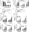
Approach and results: We, therefore, investigated whether ligands for the LXR and FXR receptors were capable of priming platelets to the activated state without stimulation by platelet agonists. Treatment of platelets with ligands for LXR and FXR converted platelets to the procoagulant state, with increases in phosphatidylserine exposure, platelet swelling, reduced membrane integrity, depolarization of the mitochondrial membrane, and microparticle release observed. Additionally, platelets also displayed features associated with coated platelets such as P-selectin exposure, fibrinogen binding, fibrin generation that is supported by increased serine protease activity, and inhibition of integrin αIIbβ3. LXR and FXR ligand-induced formation of coated platelets was found to be dependent on both reactive oxygen species and intracellular calcium mobilization, and for FXR ligands, this process was found to be dependent on cyclophilin D.
Conclusions: We conclude that treatment with LXR and FXR ligands initiates coated platelet formation, which is thought to support coagulation but results in desensitization to platelet stimuli through inhibition of αIIbβ3 consistent with their ability to inhibit platelet function and stable thrombus formation in vivo.
Keywords: bile; blood coagulation; blood platelets; calcium; cholesterol.
© 2017 The Authors.
Figures


Similar articles
-
Farnesoid X Receptor and Its Ligands Inhibit the Function of Platelets.Arterioscler Thromb Vasc Biol. 2016 Dec;36(12):2324-2333. doi: 10.1161/ATVBAHA.116.308093. Epub 2016 Oct 6. Arterioscler Thromb Vasc Biol. 2016. PMID: 27758768 Free PMC article.
-
RGD-ligand mimetic antagonists of integrin alphaIIbbeta3 paradoxically enhance GPVI-induced human platelet activation.J Thromb Haemost. 2010 Mar;8(3):567-76. doi: 10.1111/j.1538-7836.2009.03719.x. Epub 2009 Dec 11. J Thromb Haemost. 2010. PMID: 20002543
-
Platelet Activation via Shear Stress Exposure Induces a Differing Pattern of Biomarkers of Activation versus Biochemical Agonists.Thromb Haemost. 2020 May;120(5):776-792. doi: 10.1055/s-0040-1709524. Epub 2020 May 5. Thromb Haemost. 2020. PMID: 32369849 Free PMC article.
-
Platelet-based coagulation: different populations, different functions.J Thromb Haemost. 2013 Jan;11(1):2-16. doi: 10.1111/jth.12045. J Thromb Haemost. 2013. PMID: 23106920 Review.
-
Platelet-derived microparticles and the potential of glycoprotein IIb/IIIa antagonists in treating acute coronary syndrome.Tex Heart Inst J. 2009;36(2):134-9. Tex Heart Inst J. 2009. PMID: 19436807 Free PMC article. Review.
Cited by
-
Effects of sanshoamides and capsaicinoids on plasma and liver lipid metabolism in hyperlipidemic rats.Food Sci Biotechnol. 2018 Nov 15;28(2):519-528. doi: 10.1007/s10068-018-0466-2. eCollection 2019 Apr. Food Sci Biotechnol. 2018. PMID: 30956864 Free PMC article.
-
Approved LXR agonists exert unspecific effects on pancreatic β-cell function.Endocrine. 2020 Jun;68(3):526-535. doi: 10.1007/s12020-020-02241-4. Epub 2020 Mar 7. Endocrine. 2020. PMID: 32146655 Free PMC article.
-
Supramaximal calcium signaling triggers procoagulant platelet formation.Blood Adv. 2020 Jan 14;4(1):154-164. doi: 10.1182/bloodadvances.2019000182. Blood Adv. 2020. PMID: 31935287 Free PMC article.
-
Platelet Signaling Pathways and New Inhibitors.Arterioscler Thromb Vasc Biol. 2018 Apr;38(4):e28-e35. doi: 10.1161/ATVBAHA.118.310224. Arterioscler Thromb Vasc Biol. 2018. PMID: 29563117 Free PMC article. Review. No abstract available.
-
Nuclear receptors in health and disease: signaling pathways, biological functions and pharmaceutical interventions.Signal Transduct Target Ther. 2025 Jul 28;10(1):228. doi: 10.1038/s41392-025-02270-3. Signal Transduct Target Ther. 2025. PMID: 40717128 Free PMC article. Review.
References
-
- Carvalho AC, Colman RW, Lees RS. Platelet function in hyperlipoproteinemia. N Engl J Med. 1974;290:434–438. doi: 10.1056/NEJM197402212900805. - PubMed
-
- El Haouari M, Rosado JA. Platelet signalling abnormalities in patients with type 2 diabetes mellitus: a review. Blood Cells Mol Dis. 2008;41:119–123. doi: 10.1016/j.bcmd.2008.02.010. - PubMed
-
- Santilli F, Vazzana N, Liani R, Guagnano MT, Davì G. Platelet activation in obesity and metabolic syndrome. Obes Rev. 2012;13:27–42. doi: 10.1111/j.1467-789X.2011.00930.x. - PubMed
MeSH terms
Substances
Grants and funding
LinkOut - more resources
Full Text Sources
Other Literature Sources
Medical
Miscellaneous

