. 2017 Jul 4;8(27):43678-43691.
doi: 10.18632/oncotarget.18435.

Biological specificity of CDK4/6 inhibitors: dose response relationship, in vivo signaling, and composite response signature

Affiliations

Biological specificity of CDK4/6 inhibitors: dose response relationship, in vivo signaling, and composite response signature

Erik S Knudsen et al. Oncotarget. .

Abstract

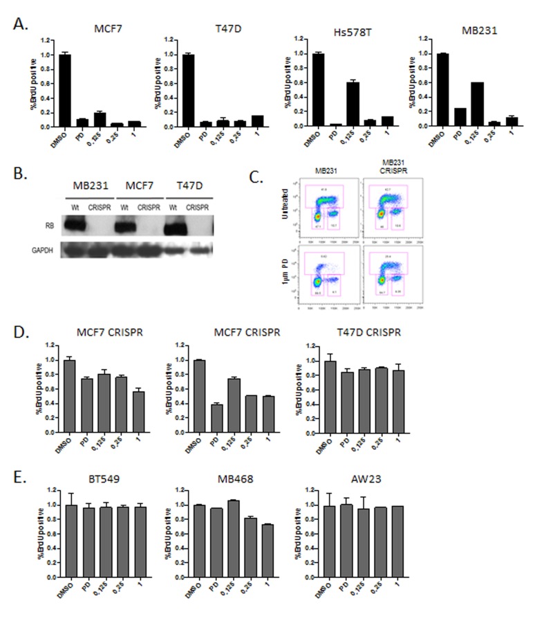
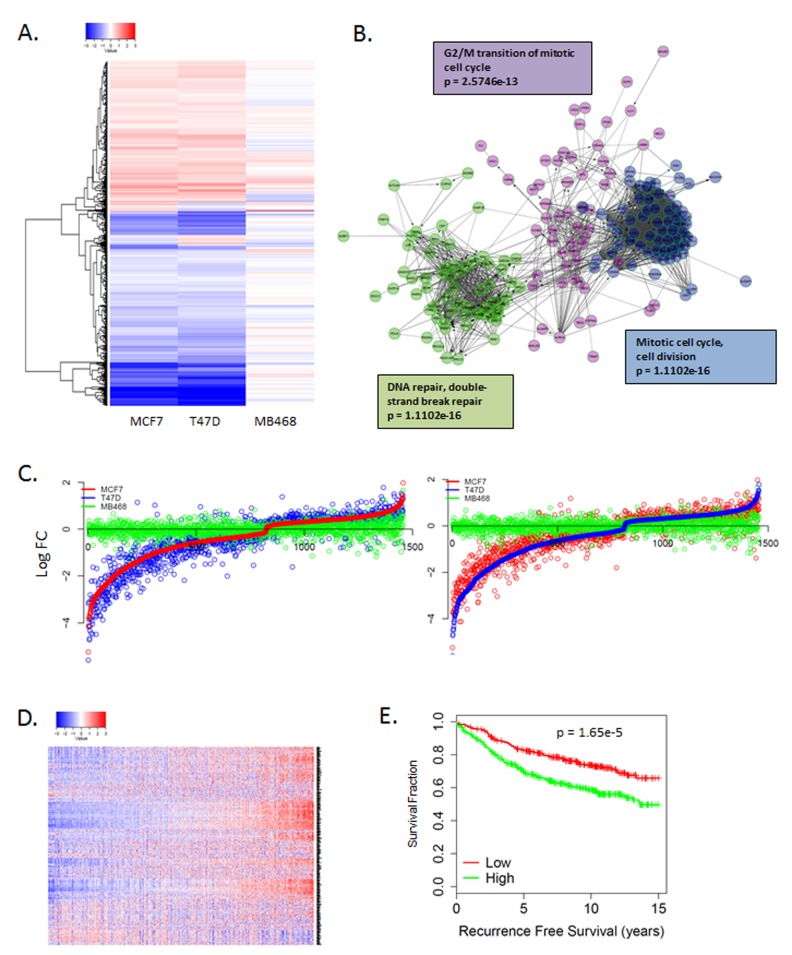
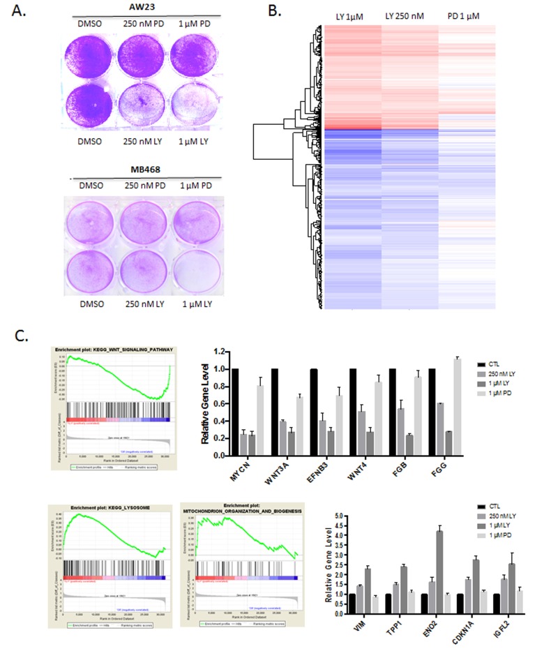
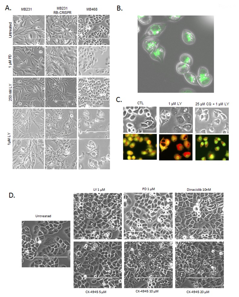
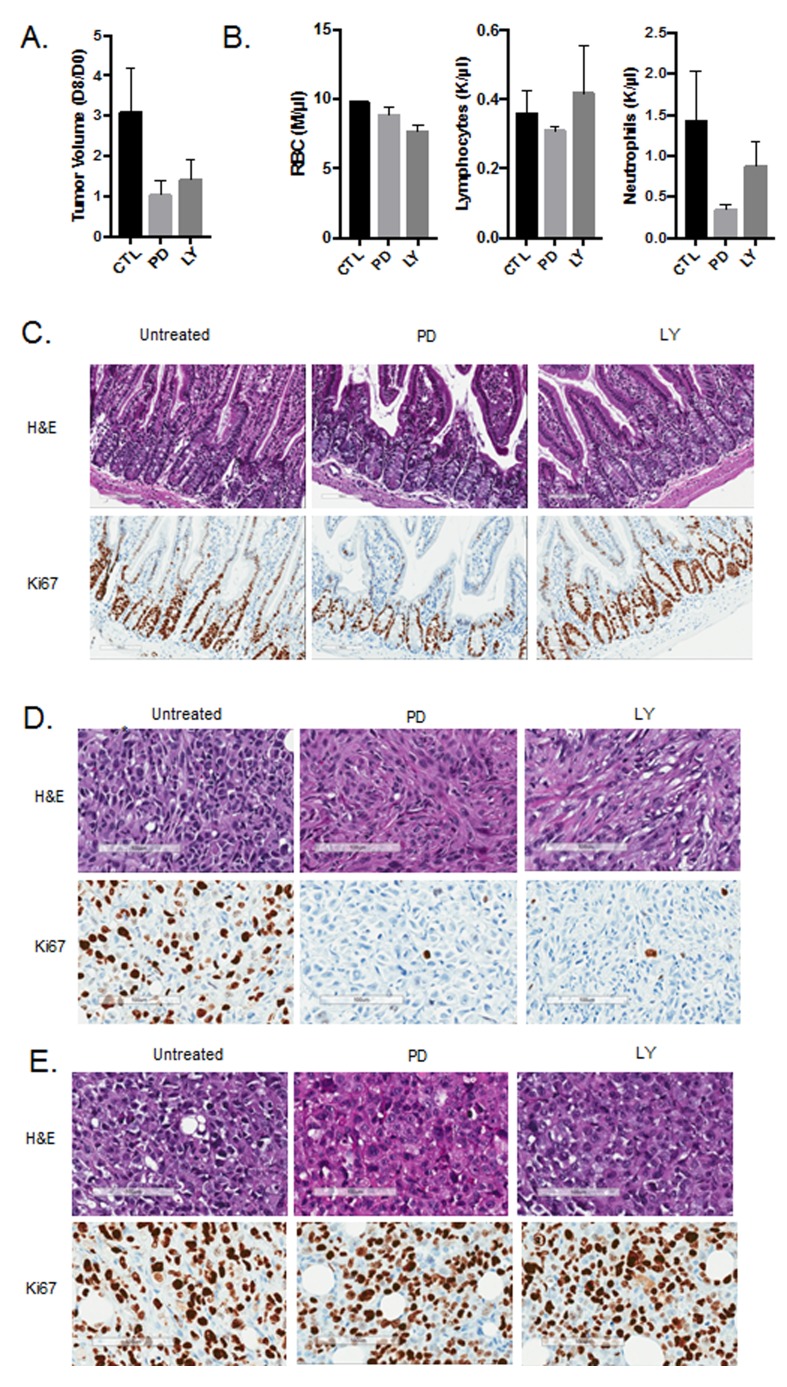
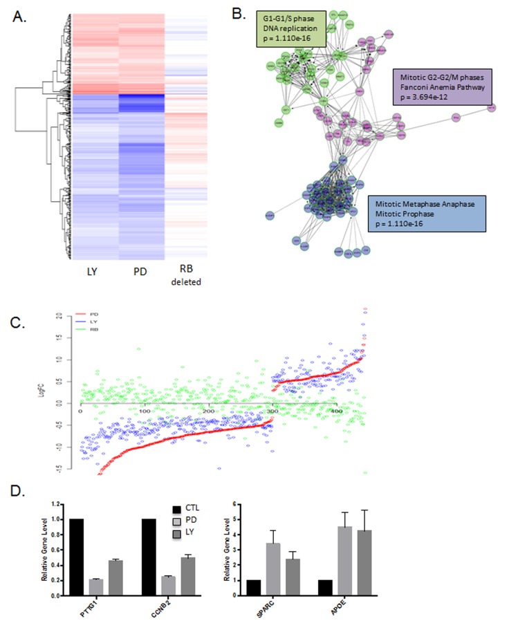
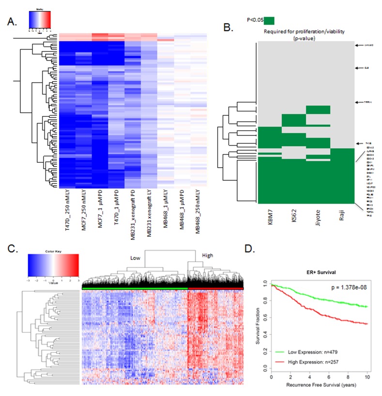
Recently developed potent and selective CDK4/6 inhibitors fall into two classes based on structure and toxicity profiles in clinical studies. One class, exemplified by palbociclib and ribociclib, exhibits neutropenia as a dose-limiting toxicity and requires discontinuous dosing. In contrast, the structurally distinct CDK4/6 inhibitor abemaciclib is dosed continuously, and has diarrhea and fatigue as dose-limiting toxicities. In preclinical models, palbociclib has been extensively studied and induces cell cycle inhibition in an RB-dependent manner. Thus far, abemaciclib has been less extensively evaluated. We found that abemaciclib cell cycle inhibitory activity is RB-dependent at clinically achievable concentrations. Abemaciclib elicited potent suppression of RB/E2F regulated genes associated with prognosis in ER-positive breast cancer. However, unlike palbociclib, at 250nM-1 µM doses abemaciclib induced cell death in RB-deficient cell lines. This response was associated with a rapidly-induced multi-vacuolar phenotype indicative of lysosomal membrane permeabilization that could be ameliorated with chloroquine. This event was not a reflection of inhibition of other CDK family members, but could be recapitulated with CBX4945 that inhibits casein and DYRK/HIPK kinases. To determine if these "off-target" features of abemaciclib were observed in vivo, mice harboring matched RB-positive and negative xenografts were treated with palbociclib and abemaciclib. In vivo, all of the apparent activity of abemaciclib was RB-dependent and strongly elicited suppression of cell cycle regulatory genes in a fashion markedly similar to palbociclib. Using gene expression data from cell lines and tumors treated with abemaciclib and palbociclib a composite signature of response to CDK4/6 inhibition was developed that included many genes that are individually required for tumor cell proliferation or viability. These data indicate that while abemaciclib and palbociclib can exert distinct biological and molecular effects, there are common gene expression features that could be broadly utilized in measuring the response to CDK4/6 inhibition.

Keywords: CDK4; E2F; abemaciclib; breast cancer; palbociclib.


Conflict of interest statement

CONFLICTS OF INTEREST

This study was supported, in part, by sponsored research funding from Eli Lilly. The work was carried out independently in an academic setting and reflects the data and interpretations of the authors.

Figures

Figure 1
Figure 1. RB-dependent cell cycle inhibitory activity
A. The indicated cell lines were treated with 1 µM palbociclib (PD) or 125 nM, 250 nM or 1 µM abemaciclib (LY). The relative BrdU incorporation was determined at 48 hours post-treatment. B. Immunoblots from the indicated cell lines developed with CRISP/Cas9 mediated deletion of RB. GAPDH is shown as a loading control. C. Representative BrdU (y-axis) vs. propidium iodide (x-axis) flow cytometry for RB-proficient and deficient models treated with palbociclib. D. The indicated cell lines were treated deleted for RB were treated with 1 µM palbociclib (PD) or 125 nM, 250 nM or 1 µM abemaciclib (LY). The relative BrdU incorporation was determined at 48 hours post-treatment. E. The indicated cell lines which are RB-deficient triple negative breast cancer models were were treated with 1 µM palbociclib (PD) or 125 nM, 250 nM or 1 µM abemaciclib (LY). The relative BrdU incorporation was determined at 48 hours post-treatment.
Figure 2
Figure 2. Unbiased gene expression response to CDK4/6 inhibition
A. Heatmap illustrating gene expression changes that occurred in MCF7 or T47D vs. MB468 cells with 250 nM abemaciclib. Data are from triplicate experiments. B. Network analysis of genes repressed by abemaciclib demonstrating key enrichment of genes regulating cell cycle transitions. C. Comparison of gene expression changes in MCF7, T47D, and MB468 cells. Log fold change is plotted on the y-axis with a given gene indicated by the dot. D. Heatmap showing the coordinate expression of abemaciclib repressed genes in a collection of 967 ER+ breast cancers. E. Kaplan-meier curve shows the association of abemaciclib repressed genes with recurrence-free survival (n = 736).
Figure 3
Figure 3. RB-independent activities of abemaciclib
A. Crystal violet staining of the indicated cell lines treated with palbociclib (PD) or abemaciclib (LY). Representative images are shown. B. Significant gene expression changes in MB468 cells are shown in the heatmap. C. Gene set enrichment analysis and expression of select induced genes with abemaciclib treatment are shown.
Figure 4
Figure 4. Defining RB-independent impacts of abemaciclib and cytotoxicity
A. The indicated cell lines were treated with the indicated doses of palbociclib (PD) and abemaciclib (LY). Representative phase contrast micrographs are shown, with the bottom row showing further magnification of the 1 µM abemaciclib treated field. B. Lysotracker green was used to localize lysosomes relative to the vacuolar structures. Representative image of MB468 cells treated with 1 µM abemaciclib are shown, merged with the phase contrast image. C. MB468 cells were treated with the indicated agents and stained with acridine orange after 6 hours of treatment. Red staining-denotes the lysomal accumulation. D. Phase contrast images of cells treated with the indicated agents.
Figure 5
Figure 5. In vivo activity of CDK4/6 inhibitors
A.Tumor volume was determined at day 0 of treatment and after 8 days of treatment. The ratio in tumor volume is shown, both abemaciclib (LY) and palbociclib (PD) significantly inhibited tumor growth (*p < 0.05). B. Complete blood counts were performed on day 8 of treatment and the total red blood cell (RBC), neutrophil, and lymphocyte counts are shown. Both abemaciclib and palbociclib treatment lead to reduced neutrophil counts. C. Immunohistochemical staining of small intestine from mice treated with the indicated agents. Ki67 staining is shown. D. Immunohistochemical staining of MB231 tumors from mice treated with the indicated agents. Ki67 staining is shown. E. Immunohistochemical staining of MB231 RB CRISPR/CAS9 deleted tumors from mice treated with the indicated agents. Ki67 staining is shown.
Figure 6
Figure 6. Gene expression responses to CDK4/6 inhibition
in vivo: A. Heatmap depicting genes that were altered by treatment with either abemaciclib (LY) or palbociclib (PD). The expression of these genes in MB231 RB CRISPR/CAS9 deleted tumors is shown for comparison. B. Network analysis of genes repressed by abemaciclib demonstrating key enrichment of genes regulating cell cycle transitions. C. Comparison of gene expression changes in LY, PD and RB deleted cells. Log fold change is plotted on the y-axis with a given gene indicated by the dot. D. Select genes that are repressed or induced by abemaciclib (LY) and palbociclib (PD).
Figure 7
Figure 7. Composite response signature for CDK4/6 inhibitors
A. Heatmap showing genes that are consistently altered with palbociclib (PD) and abemacicilb (LY) in RB-proficient models. RB-deficient MB468 model is shown as a control. B. Data plotting genes within the CDK4/6 response signature that are required for viability (p < 0.05) for the indicated cell lines. The CDK4/6 response signature genes are strongly associated with reduced viability/proliferation across all of the cell lines interrogated (two-sided test of equal proportions, p = 4.231703e-17). Select required genes are shown for reference C. Heatmap showing the relative expression of the CDK4/6 response signature across 967 ER+ breast cancer specimens. Two major clusters are identified. D. Kaplan-Meier curve dichotomized by the two major clusters observed in ER+ breast cancer specimens (log-rank p-value=1.378e-08).

Similar articles

Cited by

References

    1. Asghar U, Witkiewicz AK, Turner NC, Knudsen ES. The history and future of targeting cyclin-dependent kinases in cancer therapy. Nat Rev Drug Discov. 2015;14:130–46. doi: 10.1038/nrd4504. - DOI - PMC - PubMed
    1. O’Leary B, Finn RS, Turner NC. Treating cancer with selective CDK4/6 inhibitors. Nat Rev Clin Oncol. 2016;13:417–30. doi: 10.1038/nrclinonc.2016.26. - DOI - PubMed
    1. Sherr CJ, Beach D, Shapiro GI. Targeting CDK4 and CDK6: From Discovery to Therapy. Cancer Discov. 2016;6:353–67. doi: 10.1158/2159-8290.CD-15-0894. - DOI - PMC - PubMed
    1. Knudsen ES, Witkiewicz AK. The Strange Case of CDK4/6 Inhibitors: Mechanisms, Resistance, and Combination Strategies. Trends Cancer. 2017;3:39–55. doi: 10.1016/j.trecan.2016.11.006. - DOI - PMC - PubMed
    1. Schwartz GK, LoRusso PM, Dickson MA, Randolph SS, Shaik MN, Wilner KD, Courtney R, O’Dwyer PJ. Phase I study of PD 0332991, a cyclin-dependent kinase inhibitor, administered in 3-week cycles (Schedule 2/1) Br J Cancer. 2011;104:1862–8. doi: 10.1038/bjc.2011.177. - DOI - PMC - PubMed

MeSH terms