. 2017 Jun 15;7(1):3565.
doi: 10.1038/s41598-017-02743-8.

Phenotypic, genotypic and antigenic characterization of emerging avian reoviruses isolated from clinical cases of arthritis in broilers in Saskatchewan, Canada

Affiliations

Phenotypic, genotypic and antigenic characterization of emerging avian reoviruses isolated from clinical cases of arthritis in broilers in Saskatchewan, Canada

Lisanework E Ayalew et al. Sci Rep. .

Abstract

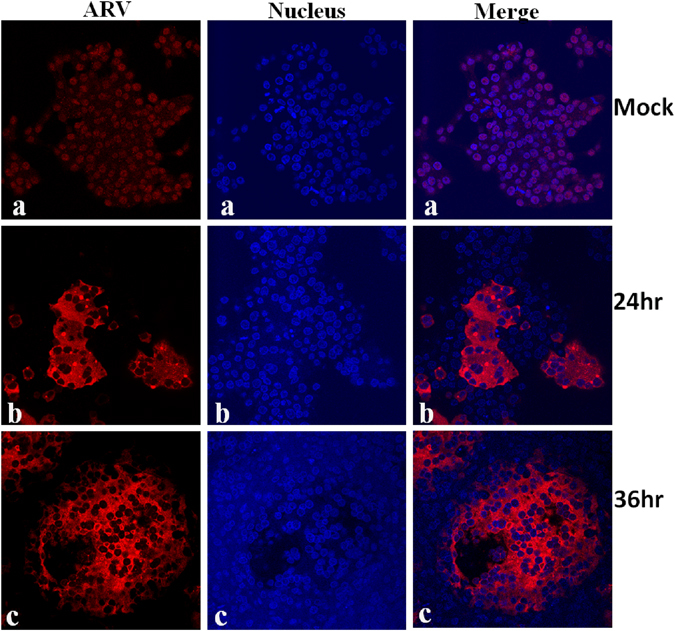
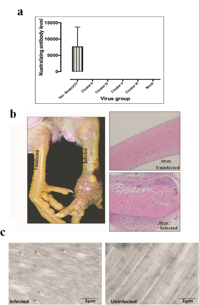
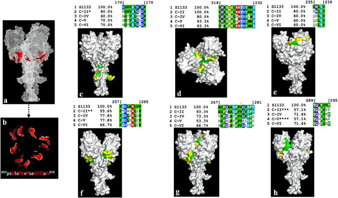
In recent years, emerging strains of pathogenic arthrogenic avian reovirus (ARV) have become a challenge to the chicken industry across USA and Canada causing significant economic impact. In this study, we characterized emerging variant ARV strains and examined their genetic and antigenic relationship with reference strains. We isolated 37 emerging variant ARV strains from tendons of broiler chickens with clinical cases of arthritis/tenosynovitis at commercial farms in Saskatchewan, Canada. Viral characterization using immunocytochemistry, gold-immunolabeling and electron microscopy revealed distinct features characteristic of ARV. Polymerase chain reaction-restriction fragment length polymorphism (PCR-RFLP) analyses of the viral Sigma C gene revealed genetic heterogeneity between the field isolates. On phylogenetic analyses, the Sigma C amino acid sequences of the isolates were clustered into four distinct genotypic groups. These ARV field strains were genetically diverse and quite distant from the vaccine and vaccine related field strains. Antibodies produced against a commercial Reo 2177 ® vaccine did not neutralize these variants. Moreover, structure based analysis of the Sigma C protein revealed significant antigenic variability between the cluster groups and the vaccine strains. To the best of our knowledge, this is the first report on the genetic, phenotypic and antigenic characterization of emerging ARVs in Canada.


Conflict of interest statement

The authors declare that they have no competing interests.

Figures

Figure 1
Figure 1
(a) The frequency distribution of ARV ELISA geometric means of 59 farms. (b) Gross and histopathological lesions in ARV infected tendon tissue, and ARV induced CPE in cell culture. (Panel a) ARV infected and non-infected tendon tissues from broiler chickens. (Panel b) Histology of normal tendon tissue. (Panel c) Lymphocytic plasmacytic infiltration in ARV infected tendon tissue with moderate number of hetrophils. (Panel d) Mock infected LMH cells. Panels (e) (f) and (g) 18, 24 and 36 hr post- infection of LMH cells with an ARV isolate, respectively.
Figure 2
Figure 2
Virus purification and TEM, as well as ultrathin sectioning of tendon tissues and immunogold labelling TEM. (a) CsCl density gradient purification of ARV virus isolate showing bands of mature virions and empty capsids. (b) Negative staining TEM of purified empty capsids. (c) Negative staining TEM of mature ARV particles. (d) and (e) Ultrathin sectioning and immunoglod labeling using anti-ARV antibody of non-infected (d) and ARV infected (e) tendon tissues after negative staining TEM.
Figure 3
Figure 3
Indirect immunostaining of mock or virus infected cells using anti ARV primary antibody and AlexaFlour 647 conjugated secondary antibody. (Panel a) mock infected LMH cells. (Panels b and c) 24 hr and 36 hr post infection of LMH cells with virus isolate, respectively. DAPI was used as a nuclear counter stain.
Figure 4
Figure 4
Polyacrylamide gel electrophoresis of the Sigma C RT-PCR products (~960 bp) following digestion with the indicated restriction enzymes. The DNA molecular weight markers are indicated on the left.
Figure 5
Figure 5
Phylogenetic tree of ARV strains based on the Sigma C sequence variability. The tree shows genetic relationship between the Sigma C protein sequence of 37 field isolates and 63 reference and variant strains isolated from around the world. The virus strains clustered into six genotyping groups (color coded). Our isolates are highlighted in bold. The tree was built by Neighbor joining method with Jukes-Cantor genetic distance model.
Figure 6
Figure 6
(a) A multiple amino acid sequence alignment of the common American vaccine strain S1133 and the 28 ARV field isolates. The alignment was made by a ClustalW alignment with BLOSUM cost matrix technique. Disagreements to the consensus sequence are highlighted. (b) Mapping of DdeI, HincII, TaqaI, BcnI and HaeIII restriction sites on Sigma C sequences of the ARV isolates (i.e. one isolate from each cluster group SK-R23 [Cluster II], SK-R16 [Cluster IV], SK-R10 [Cluster V], SK-R3 [Cluster VI]).
Figure 7
Figure 7
(a) Virus neutralization test. The neutralizing ability of antibodies produced against vaccine strain 2177 was tested against our field isolates from Cluster II, IV, V and VI. The vaccine virus strain 2177- antibody mixture and mock infected cells were used as positive and negative controls, respectively. The experiment was done in triplicates. (b) Gross and histopathological lesions of tendons from experimentally infected SPF birds. (c) Ultrathin sectioning and immunoglod labeling of tendon tissues using anti-ARV antibody from infected and non-infected SPF birds. The white arrows indicate gold labelled ARV particles.
Figure 8
Figure 8
Conserved and variable regions of predicted antigenic epitopes of the Sigma C protein shown on the modelled Sigma C carboxy terminus receptor binding domain trimeric structure. (a and b) Conserved proposed receptor binding residues. The conserved residues are highlighted in red as “cartoons”. (c to h) The conserved amino acid residues on the predicted antigenic epitopes between the S1133 vaccine strain and our isolates which grouped into different cluster “C” groups are shown in green as “surface” on the carobxy terminus receptor binding domain model structure of the Sigma C protein. The variable regions are shown in yellow. *194DV substituted by 194VM, **260M substituted by I, ***294T substituted by N or A and ****294A substituted by N or D in some cases.

Similar articles

Cited by

References

    1. Gouvea V, Schnitzer TJ. Pathogenicity of avian reoviruses: examination of six isolates and a vaccine strain. Infect Immun. 1982;38(2):731–738. - PMC - PubMed
    1. Lee LH, Wang YH, Shien JH. Serological characterization of reoviruses isolated from avian species in Taiwan. J. Chin. Soc. Vet. Sci. 1992;18:69–72.
    1. Liu HJ, Lee LH, Hsu HW, Kuo LC, Liao MH. Molecular evolution of avian reovirus: evidence for genetic diversity and reassortment of the S-class genome segments and multiple co-circulating lineages. Virology. 2003;314(1):336–49. doi: 10.1016/S0042-6822(03)00415-X. - DOI - PubMed
    1. Lu H, et al. Isolation and molecular characterization of newly emerging avian reovirus variants and novel strains in Pennsylvania, USA, 2011–2014. Sci Rep. 2015;5:14727. doi: 10.1038/srep14727. - DOI - PMC - PubMed
    1. Schnitzer TJ, Ramos T, Gouvea V. Avian Reovirus Polypeptides: Analysis of Intracellular Virus-Specified Products, Virions, Top Component, and Cores. J Virol. 1982;43(3):1006–1014. - PMC - PubMed

Publication types

MeSH terms