. 2017 May 31:8:656.
doi: 10.3389/fimmu.2017.00656. eCollection 2017.

Vitamin D Counteracts Mycobacterium tuberculosis-Induced Cathelicidin Downregulation in Dendritic Cells and Allows Th1 Differentiation and IFNγ Secretion

Affiliations

Vitamin D Counteracts Mycobacterium tuberculosis-Induced Cathelicidin Downregulation in Dendritic Cells and Allows Th1 Differentiation and IFNγ Secretion

Anna K O Rode et al. Front Immunol. .

Abstract

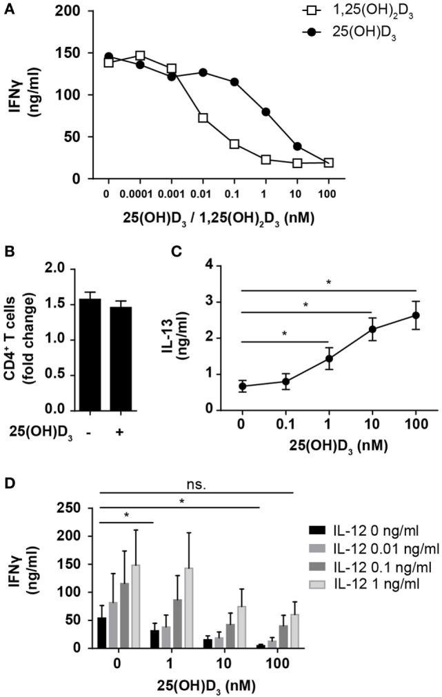
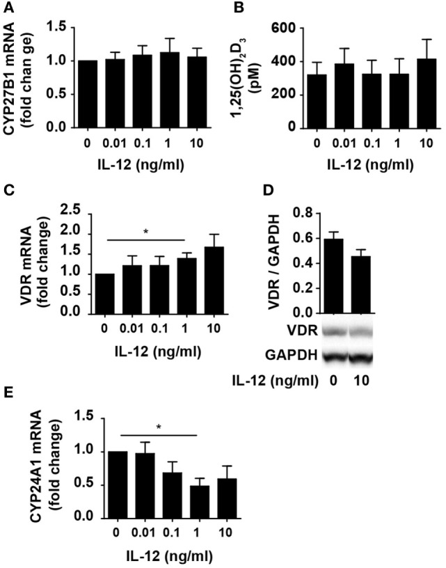
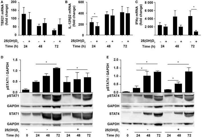
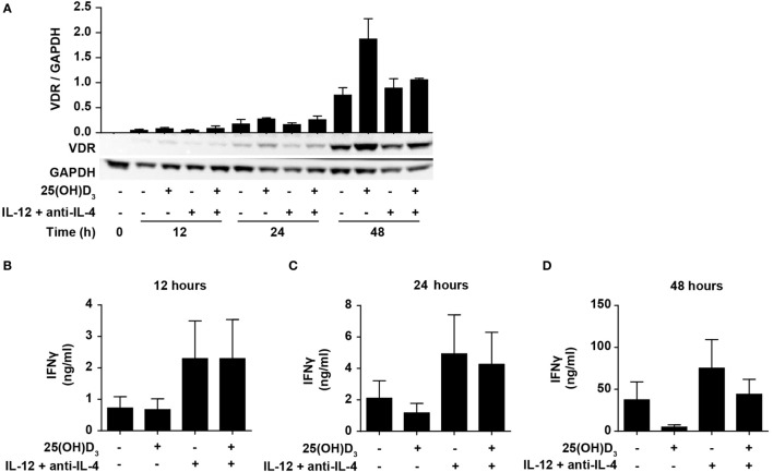
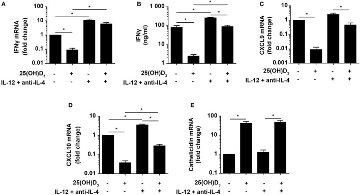
Tuberculosis (TB) presents a serious health problem with approximately one-third of the world's population infected with Mycobacterium tuberculosis in a latent state. Experience from the pre-antibiotic era and more recent clinical studies have established a beneficial role of sunlight and vitamin D in patients with TB. At the same time, experimental data have shown that Th1 cells through production of IFNγ are crucial for cathelicidin release by macrophages, bacterial killing, and containment of M. tuberculosis in granulomas. Paradoxically, vitamin D has repeatedly been ascribed an immune-suppressive function inhibiting Th1 differentiation and production of IFNγ in T cells. The aim of this study was to investigate this apparent paradox. We studied naïve human CD4+ T cells activated either with CD3 and CD28 antibodies or with allogeneic dendritic cells (DC) stimulated with heat-killed M. tuberculosis (HKMT) or purified toll-like receptor (TLR) ligands. We show that vitamin D does not block differentiation of human CD4+ T cells to Th1 cells and that interleukin (IL)-12 partially counteracts vitamin D-mediated inhibition of IFNγ production promoting production of equal amounts of IFNγ in Th1 cells in the presence of vitamin D as in T cells activated in the absence of vitamin D and IL-12. Furthermore, we show that HKMT and TLR2 ligands strongly downregulate cathelicidin expression in DC and that vitamin D counteracts this by upregulating cathelicidin expression. In conclusion, we demonstrate that vitamin D counteracts M. tuberculosis-induced cathelicidin downregulation and allows Th1 differentiation and IFNγ secretion.

Keywords: IFNγ; T cells; Th1; cathelicidin; dendritic cells; tuberculosis; vitamin D.


Figures

Figure 1
Figure 1
IL-12 counteracts vitamin D-mediated inhibition of IFNγ production in CD4+ T cells. (A) IFNγ concentration in the supernatants of T cells activated for 72 h in the presence of the indicated concentrations of 25(OH)D3 or 1,25(OH)2D3. Representative graphs from two independent experiments with three donors are shown. (B) Number of CD4+ T cells in the cell cultures after activation for 72 h in the presence or absence of 100 nM 25(OH)D3 relative to initial cell number (mean + SEM, n = 12). (C) IL-13 concentration in the supernatant of T cells activated for 72 h in the presence of the indicated concentrations of 25(OH)D3 (mean ± SEM, n = 6). (D) IFNγ concentration in the supernatants of T cells activated for 72 h in the presence of the indicated concentrations of 25(OH)D3 and IL-12 (mean + SEM, n ≥ 4).
Figure 2
Figure 2
IL-12 does not inhibit 1,25(OH)2D3 production or VDR expression and function in CD4+ T cells. Relative CYP27B1 (A), VDR (C), and CYP24A1 (E) expression in T cells activated for 72 h in the presence of 100 nM 25(OH)D3 and the indicated concentration of IL-12. Data are normalized to activated T cells incubated with 100 nM 25(OH)D3 in the absence of IL-12 (mean + SEM, n ≥ 6). (B) 1,25(OH)2D3 production in T cells activated for 72 h in the presence of 100 nM 25(OH)D3 and the indicated concentration of IL-12 (mean + SEM, n ≥ 4). (D) Representative Western blot (lower panel) and quantification (upper panel) of VDR with GAPDH as loading control from T cells activated for 72 h in the presence of 100 nM 25(OH)D3 and in the presence or absence of 10 ng/ml IL-12 (mean + SEM, n = 4). Western blots including protein ladder are shown in the Figure S1 in Supplementary Material.
Figure 3
Figure 3
Vitamin D does not prevent Th1 differentiation. Relative TBX21 (A), IL-12Rβ2 (B), and IFNγ (C) expression in T cells activated for 24, 48, or 72 h in Th1-polarizing medium in the presence or absence of 100 nM 25(OH)D3. Data are normalized to unstimulated T cells (mean + SEM, n = 6). Representative Western blots (lower panel) and quantification (upper panel) of phosphorylated STAT1 and total STAT1 (D) and phosphorylated STAT4 and total STAT4 (E) with GAPDH as loading control from T cells activated for 0, 24, 48, and 72 h in Th1-polarizing medium in the presence or absence of 100 nM 25(OH)D3. Western blots including protein ladder are shown in the Figure S2 in Supplementary Material.
Figure 4
Figure 4
IL-12-mediated signaling is initiated before vitamin D signaling. (A) Representative Western blots (lower panel) and quantification (upper panel) of VDR with GAPDH as loading control from T cells activated for 0, 12, 24, and 48 h in the presence or absence of 100 nM 25(OH)D3 and Th1-polarizing medium (IL-12 + anti-IL-4) (mean + SEM, n = 2). IFNγ concentration in the supernatants of T cells activated for 12 h (B), 24 h (C), or 48 h (D) in the presence or absence of 100 nM 25(OH)D3 and Th1-polarizing medium (mean + SEM, n = 4). Western blots including protein ladder are shown in the Figure S3 in Supplementary Material.
Figure 5
Figure 5
Vitamin D is required for concomitant production of IFNγ and cathelicidin in DC–T cell cultures. Relative expression (A) and production (B) of IFNγ and relative CXCL9 (C), CXCL10 (D), and cathelicidin (E) expression in DC–T cell co-cultures incubated in the presence or absence of 100 nM 25(OH)D3 and Th1-polarizing medium (IL-12 + anti-IL-4). Data are normalized to the values obtained from co-cultures incubated in the absence of 25(OH)D3 and Th1-polarizing medium (mean + SEM, n = 4).
Figure 6
Figure 6
Vitamin D counteracts the inhibitory effect of M. tuberculosis on cathelicidin production in DC. Relative cathelicidin (A), CYP27B1 (B), VDR (D), and CYP24A1 (E) expression and 1,25(OH)2D3 production (C) in untreated DC (no TLR ligand) or DC treated with HKMT or Pam3CSK4 and subsequently incubated for 24 h in the absence or presence of 100 nM 25(OH)D3 and 10 ng/ml IFNγ. Data are normalized to the values obtained from untreated DC in the absence of 25(OH)D3 and IFNγ (mean + SEM, n ≥ 3), *indicates p ≤ 0.05. Relative cathelicidin (F) and CYP24A1 (G) expression in untreated DC (no TLR ligand) or DC treated with HKMT or Pam3CSK4 incubated for 24 h with the indicated concentrations of 1,25(OH)2D3. Data are normalized to untreated DC in the absence of 1,25(OH)2D3 (mean ± SEM, n ≥ 3), * indicates p ≤ 0.05 for no TLR ligand untreated versus 1,25(OH)2D3 treated, # indicates p ≤ 0.05 for HKMT versus no TLR ligand, ¤ indicates p ≤ 0.05 for Pam3CSK4 versus no TLR ligand.

References

    1. WHO. Global Tuberculosis Report 2016. (2016). Available from: www.who.int/tb/publications/global_report/en/
    1. Nunes-Alves C, Booty MG, Carpenter SM, Jayaraman P, Rothchild AC, Behar SM. In search of a new paradigm for protective immunity to TB. Nat Rev Microbiol (2014) 12:289–99. 10.1038/nrmicro3230 - DOI - PMC - PubMed
    1. Getahun H, Matteelli A, Chaisson RE, Raviglione M. Latent Mycobacterium tuberculosis infection. N Engl J Med (2015) 372:2127–35. 10.1056/NEJMra1405427 - DOI - PubMed
    1. Srivastava S, Ernst JD, Desvignes L. Beyond macrophages: the diversity of mononuclear cells in tuberculosis. Immunol Rev (2014) 262:179–92. 10.1111/imr.12217 - DOI - PMC - PubMed
    1. Humphreys IR, Stewart GR, Turner DJ, Patel J, Karamanou D, Snelgrove RJ, et al. A role for dendritic cells in the dissemination of mycobacterial infection. Microbes Infect (2006) 8:1339–46. 10.1016/j.micinf.2005.12.023 - DOI - PubMed