. 2017:2017:5821706.
doi: 10.1155/2017/5821706. Epub 2017 May 23.

Periplaneta americana Extracts Promote Skin Wound Healing via Nuclear Factor Kappa B Canonical Pathway and Extracellular Signal-Regulated Kinase Signaling

Affiliations

Periplaneta americana Extracts Promote Skin Wound Healing via Nuclear Factor Kappa B Canonical Pathway and Extracellular Signal-Regulated Kinase Signaling

Qin Song et al. Evid Based Complement Alternat Med. 2017.

Abstract

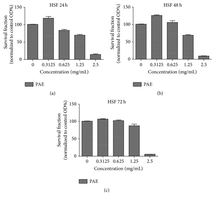
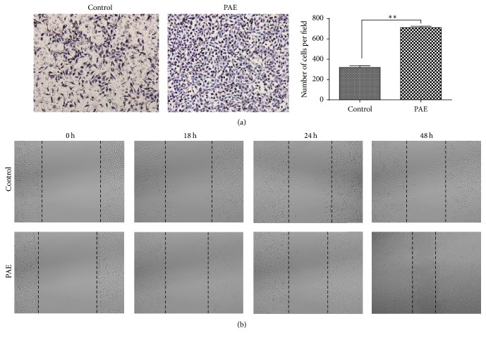
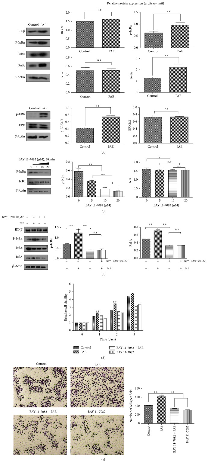
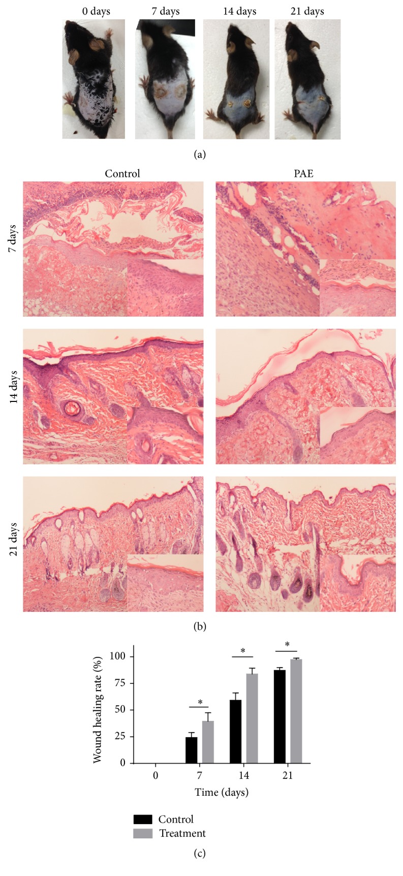
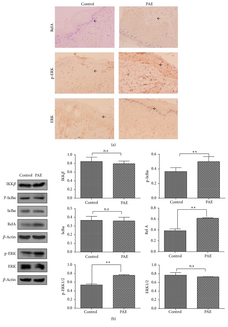
Periplaneta americana extracts (PAEs) exhibit wound healing properties. However, the underlying molecular mechanisms are not well understood. Here, we treated human skin fibroblasts (HSF) with PAE and the proliferation was determined by 3-(4,5-dimethylthiazol-2-yl)-2,5-diphenyltetrazolium bromide (MTT) assay. The wound healing and transwell migration assays were used to detect cell migration. Nuclear factor kappa B (NF-κB) and extracellular signal-regulated kinase (ERK) pathways were analyzed by Western blot (WB). Immunofluorescence staining was used to detect the key molecular localization in the cells. The results showed that PAE enhanced the proliferation and migration of HSF cells. The expression and activation of key proteins such as RelA and p-ERK were increased in NF-κB and ERK pathways followed by nuclear translocation. In vivo, both WB and immunohistochemical (IHC) staining showed that PAE enhanced p-IκBα and p-ERK activation and the nuclear translocation of RelA. Our study suggests that the protective function of PAE is mediated via enhanced NF-κB and ERK signaling.


Figures

Figure 1
Figure 1
Chromatographic separation of PAE. (a) HPLC chromatogram of authentic standards tested. (b) HPLC chromatogram of PAE at 254 nm: peak 1, uracil (1.140 mg/g); peak 2, hypoxanthine (4.257 mg/g); and peak 3, inosine (8.158 mg/g). Identification was based on retention time and UV spectra compared with commercial standards.
Figure 2
Figure 2
MTT assay using different doses of PAE on HSF at different time points. (a) The survival fraction of different doses of extract on HSF at 24 h (normalized to control group); (b) the survival fraction of different doses of extract on HSF at 48 h (normalized to control group); (c) the survival fraction of different doses of extract on HSF at 72 h (normalized to control group).
Figure 3
Figure 3
Effect of PAE on cell migration in HSF cells. The differentially cultured HSF (0.3125 mg/mL) for 48 h. (a) Transwell migration assay: images obtained at 24 h after HSF were incubated in Milicells 24 h (left); the migratory cells per visual field (100x) were counted and expressed as the average numbers. Data represent mean ± SD. ∗∗p < 0.01 versus control group (right). (b) Cell scratch test: images obtained at 0, 18, 24, and 48 h after scratch formation.
Figure 4
Figure 4
PAE affected NF-κB canonical and ERK pathways. HSF cells were differentially cultured for 48 h, and the protein expression of (a) NF-κB canonical pathway and ERK pathway varied; (b) NF-κB canonical pathway proteins in the presence of BAY 11-7082; (c) NF-κB pathway expression in the presence of BAY 11-7082 and PAE. Actin was used as a loading control. Cells were incubated with a medium containing PAE at the indicated concentrations for 48 h after pretreatment with BAY 11-7082 (10 μM) for 30 min. Cell proliferation (d) was determined by MTT and (e) migration was tested by transwell assay. Data represent mean ± SD. p < 0.05; ∗∗p < 0.01 versus control group.
Figure 5
Figure 5
Immunofluorescence analysis of the spatial localization of the NF-κB canonical and ERK pathways. Cells were treated with PAE for 48 h, fixed, and incubated to determine the immunofluorescence. The antibodies against (a) RelA, (b) p-ERK, and (c) ERK presented green fluorescence, whereas a blue fluorescent signal was generated by DAPI staining of the cell nuclei. Areas of overlap between the green and the blue fluorescence appeared as merged images.
Figure 6
Figure 6
PAE affected wound healing in cutaneous thermal burns in vivo. In the C57 mouse model of thermal injury, the left wound of the dorsal flank was treated with PAE (5 mg/mL) while the right side treated with normal saline served as the daily control. (a) Representative images of thermal injury on days 0, 7, 14, and 21; (b) histological HE staining of differentially treated tissues on days 0, 7, 14, and 21; (c) healing rate of thermal wound. p < 0.05 versus control group.
Figure 7
Figure 7
NF-κB canonical pathway and ERK pathway activities in vivo. (a) Molecular expression of NF-κB canonical pathway and ERK pathway in PAE-treated and control tissues was assayed by IHC, and the representative images are shown (400x). (b) Western blot of protein expression in NF-κB canonical and ERK pathways. Actin was used as a loading control. Data represent mean ± SD. ∗∗p < 0.01 versus control group.

References

    1. Shi H. X., Lin C., Lin B. B., et al. The anti-scar effects of basic fibroblast growth factor on the wound repair in vitro and in vivo. PLoS ONE. 2013;8(4) doi: 10.1371/journal.pone.0059966.e59966 - DOI - PMC - PubMed
    1. Li M., Zhao Y., Hao H., et al. Mesenchymal stem cell-conditioned medium improves the proliferation and migration of keratinocytes in a diabetes-like microenvironment. The International Journal of Lower Extremity Wounds. 2015;14(1):73–86. doi: 10.1177/1534734615569053. - DOI - PubMed
    1. Chen X., Ran X., Sun R., et al. Protective effect of an extract from Periplaneta americana on hematopoiesis in irradiated rats. International Journal of Radiation Biology. 2009;85(7):607–613. doi: 10.1080/09553000902985110. - DOI - PubMed
    1. Brown A. E., France R. M., Grossman S. H. Purification and characterization of arginine kinase from the American cockroach (Periplaneta americana) Archives of Insect Biochemistry and Physiology. 2004;56(2):51–60. doi: 10.1002/arch.10143. - DOI - PubMed
    1. Jeong K. Y., Hwang H., Lee J., et al. Allergenic characterization of tropomyosin from the dusky brown cockroach, periplaneta fuliginosa. Clinical and Vaccine Immunology. 2004;11(4):680–685. doi: 10.1128/CDLI.11.4.680-685.2004. - DOI - PMC - PubMed

LinkOut - more resources