Review
. 2015 Oct 15:1:15.
doi: 10.1186/s40738-015-0008-z. eCollection 2015.

Incidence, diagnosis and management of tubal and nontubal ectopic pregnancies: a review

Affiliations
Review

Incidence, diagnosis and management of tubal and nontubal ectopic pregnancies: a review

Danielle M Panelli et al. Fertil Res Pract. .

Abstract

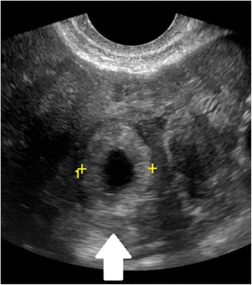
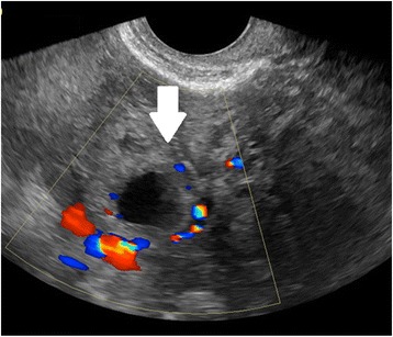
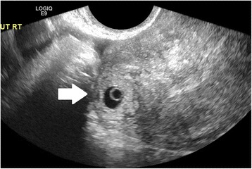
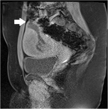
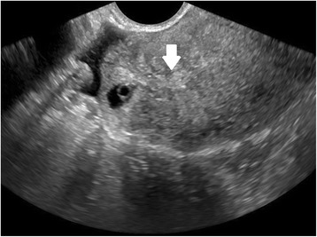
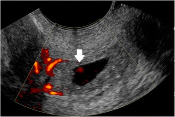
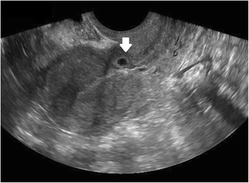
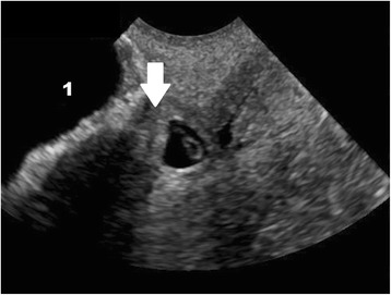
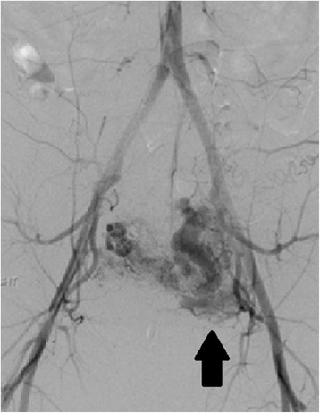
Background: Ectopic pregnancy is a potentially life-threatening condition occurring in 1-2 % of all pregnancies. The most common ectopic implantation site is the fallopian tube, though 10 % of ectopic pregnancies implant in the cervix, ovary, myometrium, interstitial portion of the fallopian tube, abdominal cavity or within a cesarean section scar.

Findings: Diagnosis involves a combination of clinical symptoms, serology, and ultrasound. Medical management is a safe and effective option in most clinically stable patients. Patients who have failed medical management, are ineligible, or present with ruptured ectopic pregnancy or heterotopic pregnancy are most often managed with excision by laparoscopy or, less commonly, laparotomy. Management of nontubal ectopic pregnancies may involve medical or surgical treatment, or a combination, as dictated by ectopic pregnancy location and the patient's clinical stability. Following tubal ectopic pregnancy, the rate of subsequent intrauterine pregnancy is high and independent of treatment modality.

Conclusion: This review describes the incidence, risk factors, diagnosis, and management of tubal and non-tubal ectopic and heterotopic pregnancies, and reviews the existing data regarding recurrence and future fertility.

Keywords: Ectopic pregnancy; Heterotopic pregnancy; Nontubal ectopic pregnancy.


Figures

Fig. 1
Fig. 1
Tubal ectopic pregnancy by transvaginal ultrasound. The arrow indicates the ectopic gestation with a surrounding hyperechoic ring, called the ‘bagel’ or ‘tubal’ sign
Fig. 2
Fig. 2
Tubal ectopic pregnancy by transvaginal ultrasound. The arrow indicates the ectopic gestation with circumferential Doppler flow, called the “Ring of Fire”
Fig. 3
Fig. 3
Interstitial ectopic pregnancy by transvaginal ultrasound. The arrow indicates thin (<5 mm) myometrium overlying the ectopic pregnancy. This finding by ultrasound, in combination with the lateral location of the gestation, has a reported specificity of 88-93 % but a sensitivity of just 40 % [101].
Fig. 4
Fig. 4
Interstitial ectopic pregnancy by magnetic resonance imaging, T1 weighted. The arrow indicates thin (<5 mm) myometrium overlying the ectopic pregnancy. In a stable patient, MRI may be useful in the confirmation of interstitial pregnancy location
Fig. 5
Fig. 5
Interstitial ectopic pregnancy by transvaginal ultrasound. The arrow indicates the ‘interstitial line,’ extending from the endometrium to the cornua, abutting the suspicious mass
Fig. 6
Fig. 6
Cervical ectopic pregnancy by transvaginal ultrasound. Doppler shows circumferential flow. The arrow indicates Doppler flow inside the gestational sac, associated with the embryo. Such Doppler flow will not be found in a spontaneous abortion, which may slide down into a similar position at the cervix
Fig. 7
Fig. 7
Cesarean scar ectopic pregnancy by transvaginal ultrasound. The arrow shows the gestational sac implanted in the region of the cesarean scar, clearly outside the endometrial canal
Fig. 8
Fig. 8
Cesarean scar ectopic pregnancy by transvaginal ultrasound. The arrow indicates the thin myometrium (3 mm) between the bladder (indicated with the number 1) and the gestational sac
Fig. 9
Fig. 9
Left uterine artery arterio-venous malformation (AVM) by pelvic angiogram. This patient had undergone an uncomplicated ultrasound-guided D&C for a 10 week size cesarean scar ectopic pregnancy 2 months prior to presentation with vaginal bleeding and diagnosis of a left uterine artery AVM (arrow). The AVM was embolized with coils, but the patient required emergent hysterectomy for hemorrhage

Similar articles

Cited by

References

    1. Creanga AA, Shapiro-Mendoza CK, Bish CL, Zane S, Berg CJ, Callaghan WM. Trends in ectopic pregnancy mortality in the United States: 1980–2007. Obstet Gynecol. 2011;117:837–843. doi: 10.1097/AOG.0b013e3182113c10. - DOI - PubMed
    1. Arleo EK, DeFilippis EM. Cornual, interstitial, and angular pregnancies: clarifying the terms and a review of the literature. Clin Imaging. 2014;38:763–770. doi: 10.1016/j.clinimag.2014.04.002. - DOI - PubMed
    1. Hoffman BL, Schorge JO, Schaffer JI, Halvorson LM, Bradshaw KD, Cunningham F, et al. Chapter 7. Ectopic pregnancy. In: Hoffman BL, Schorge JO, Schaffer JI, Halvorson LM, Bradshaw KD, Cunningham F, Calver LE, et al., editors. Williams gynecology. 2. New York: McGraw-Hill; 2012.
    1. Lau S, Tulandi T. Conservative medical and surgical management of interstitial ectopic pregnancy. Fertil Steril. 1999;72:207–215. doi: 10.1016/S0015-0282(99)00242-3. - DOI - PubMed
    1. Barnhart K. Ectopic pregnancy. N Engl J Med. 2009;361:379–387. doi: 10.1056/NEJMcp0810384. - DOI - PubMed

LinkOut - more resources