

Suprascapular Nerve Entrapment Caused by Protrusion of an Intraosseous Ganglion of the Glenoid into the Spinoglenoid Notch: A Rare Cause of Posterior Shoulder Pain
- PMID: 28620557
- PMCID: PMC5460384
- DOI: 10.1155/2017/1704697
Suprascapular Nerve Entrapment Caused by Protrusion of an Intraosseous Ganglion of the Glenoid into the Spinoglenoid Notch: A Rare Cause of Posterior Shoulder Pain
Abstract
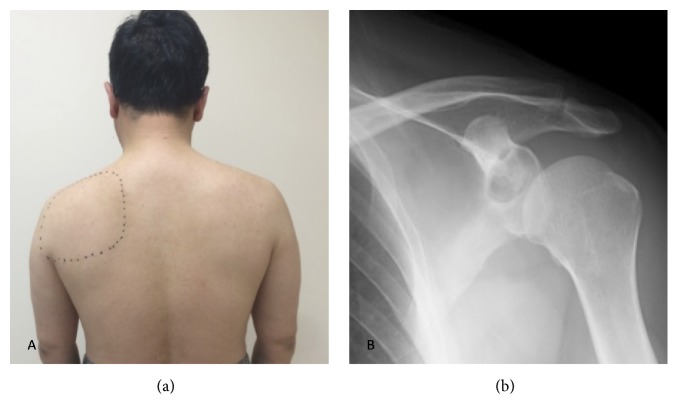
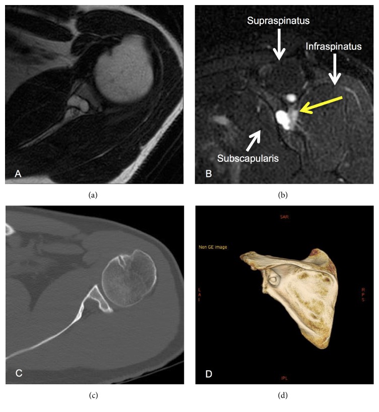
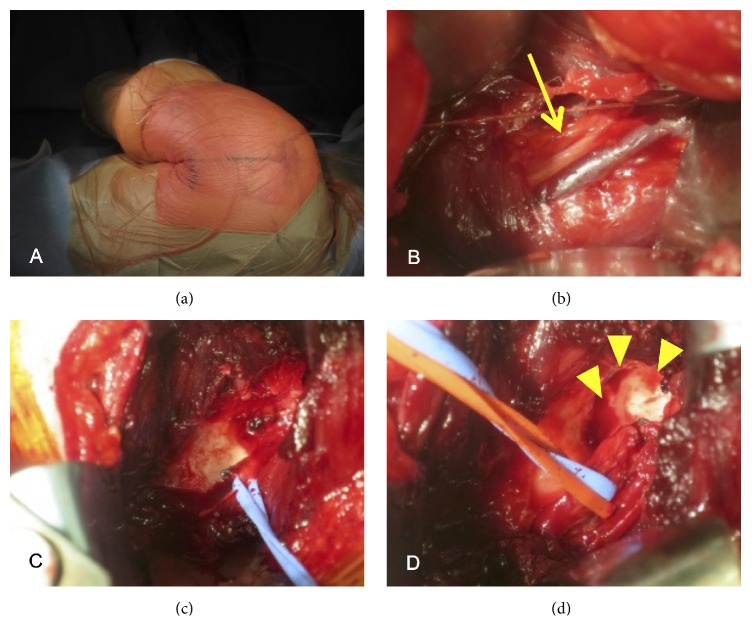
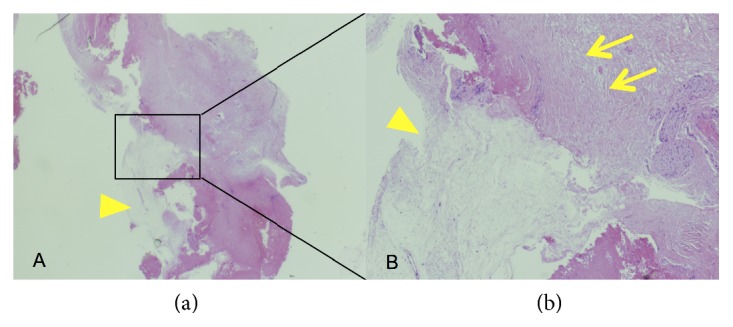
We describe a case of suprascapular nerve entrapment caused by protrusion of an intraosseous ganglion of the glenoid into the spinoglenoid notch. A 47-year-old man with left shoulder pain developed an intraosseous cyst in the left glenoid, which came into contact with the suprascapular nerve. The area at which the patient experienced spontaneous shoulder pain was innervated by the suprascapular nerve, and 1% xylocaine injection into the spinoglenoid notch under ultrasonographic guidance relieved the pain. Therefore, we concluded that the protrusion of an intraosseous cyst of the glenoid into the spinoglenoid notch was a cause of the pain, and performed curettage. Consequently, the shoulder pain was resolved promptly without suprascapular nerve complications, and the cyst was histologically diagnosed as an intraosseous ganglion. This case demonstrated that the intraosseous ganglion of the glenoid was a benign lesion but could be a cause of suprascapular nerve entrapment syndrome. Curettage is a useful treatment option for a ganglion inside bone and very close to the suprascapular nerve.
Figures
Similar articles
-
Intraosseous Ganglion Protruding Into the Spinoglenoid Notch With Suprascapular Nerve Entrapment: A Case Report.Cureus. 2024 Nov 30;16(11):e74863. doi: 10.7759/cureus.74863. eCollection 2024 Nov. Cureus. 2024. PMID: 39737318 Free PMC article.
-
Suprascapular nerve entrapment caused by a large hematoma of the scapula: a case report.BMC Musculoskelet Disord. 2023 Jul 19;24(1):589. doi: 10.1186/s12891-023-06723-0. BMC Musculoskelet Disord. 2023. PMID: 37468872 Free PMC article.
-
Suprascapular nerve entrapment caused by an intraosseous ganglion of the scapula: A case report.Medicine (Baltimore). 2017 Jun;96(24):e7167. doi: 10.1097/MD.0000000000007167. Medicine (Baltimore). 2017. PMID: 28614252 Free PMC article.
-
[Ganglion cyst at the spinoglenoid notch. A case report and literature review].Rev Med Interne. 2005 Apr;26(4):335-8. doi: 10.1016/j.revmed.2004.12.006. Epub 2005 Jan 26. Rev Med Interne. 2005. PMID: 15820571 Review. French.
-
Suprascapular nerve neuropathy secondary to spinoglenoid notch ganglion cyst: case reports and review of literature.Ann Acad Med Singap. 2007 Dec;36(12):1032-5. Ann Acad Med Singap. 2007. PMID: 18185886 Review.
Cited by
-
Intraosseous Ganglion Protruding Into the Spinoglenoid Notch With Suprascapular Nerve Entrapment: A Case Report.Cureus. 2024 Nov 30;16(11):e74863. doi: 10.7759/cureus.74863. eCollection 2024 Nov. Cureus. 2024. PMID: 39737318 Free PMC article.
-
Shoulder Agony: A Painful Glenoid Cyst.Cureus. 2023 Jan 12;15(1):e33726. doi: 10.7759/cureus.33726. eCollection 2023 Jan. Cureus. 2023. PMID: 36788828 Free PMC article.
-
Suprascapular nerve entrapment caused by a large hematoma of the scapula: a case report.BMC Musculoskelet Disord. 2023 Jul 19;24(1):589. doi: 10.1186/s12891-023-06723-0. BMC Musculoskelet Disord. 2023. PMID: 37468872 Free PMC article.
References
-
- Urayama M., Itoi E., Watanabe H., Sato K., Kamei J. Intraosseous ganglion of the glenoid. Orthopedics. 1999;22(7):705–706. - PubMed
Publication types
LinkOut - more resources
Full Text Sources
Other Literature Sources
