. 2017 Jun 16:8:15847.
doi: 10.1038/ncomms15847.

Structural insights into the function of ZRANB3 in replication stress response

Affiliations

Structural insights into the function of ZRANB3 in replication stress response

Marek Sebesta et al. Nat Commun. .

Abstract

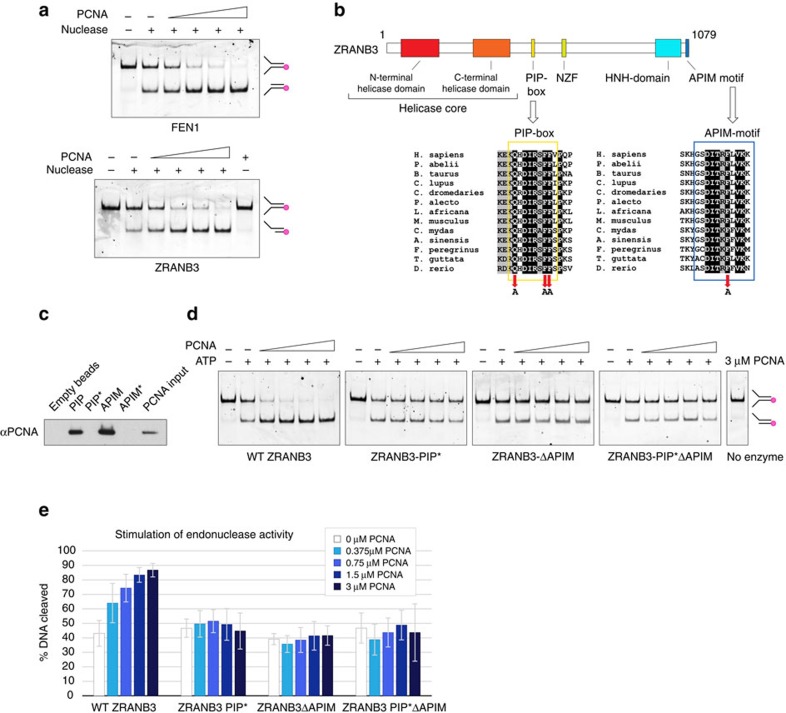
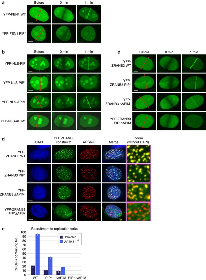
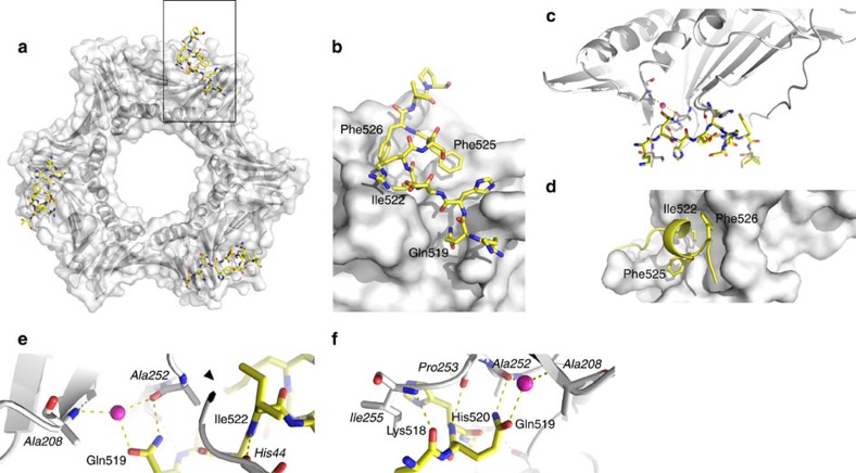
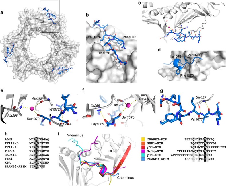
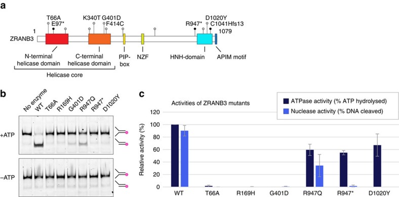
Strategies to resolve replication blocks are critical for the maintenance of genome stability. Among the factors implicated in the replication stress response is the ATP-dependent endonuclease ZRANB3. Here, we present the structure of the ZRANB3 HNH (His-Asn-His) endonuclease domain and provide a detailed analysis of its activity. We further define PCNA as a key regulator of ZRANB3 function, which recruits ZRANB3 to stalled replication forks and stimulates its endonuclease activity. Finally, we present the co-crystal structures of PCNA with two specific motifs in ZRANB3: the PIP box and the APIM motif. Our data provide important structural insights into the PCNA-APIM interaction, and reveal unexpected similarities between the PIP box and the APIM motif. We propose that PCNA and ATP-dependency serve as a multi-layered regulatory mechanism that modulates ZRANB3 activity at replication forks. Importantly, our findings allow us to interpret the functional significance of cancer associated ZRANB3 mutations.


Conflict of interest statement

The authors declare no competing financial interests.

Figures

Figure 1
Figure 1. Crystal structure of the ZRANB3 HNH domain.
(a) Crystal structure of the ZRANB3 HNH domain (residues 948–1,067). Secondary structure elements are annotated (helices α1–α8 and β-strands β1–β2). A bound zinc ion is shown as an orange sphere, with its four coordinating cysteine residues shown as sticks. The ZRANB3-specific helical domain is shown in blue. (b) Top scoring structural homologues of the ZRANB3 HNH domain identified by DALI search. (c) Sequence alignment of the HNH domains from eukaryotic ZRANB3 proteins, bacterial self-standing HNH nucleases and bacterial ZRANB3-like proteins. Secondary structure elements are shown for human ZRANB3 HNH domain only. Targeted mutation sites are indicated by red (basic residues) and blue (active site residues) arrows; residues involved in zinc ion coordination are indicated by yellow arrows; T4 endonuclease VII active site residues are indicated by purple arrows. (d) Overlay of the ZRANB3 HNH (white) with the structures of T4 endonuclease VII (yellow) and Geobacter metallireducens HNH nicking endonuclease (orange). Non-catalytic zinc ions are shown as orange spheres. (e) Nuclease assay with full-length ZRANB3 and the two mutants containing deletions of the ZRANB3-specific helical domain (ZRANB3-Δ975–1013 and ZRANB3-Δ972–1010). DNA substrate (splayed DNA duplex fluorescently labelled at the 5′ end) and the resulting product of the nucleolytic cleavage (fluorescently labelled DNA duplex with 5′ overhang) are indicated in the picture. Shown are reactions in the presence and absence of ATP. Reactions were analysed by native polyacrylamide gel electrophoresis.
Figure 2
Figure 2. DNA binding surface.
(a) Electrostatic surface potential (blue, positive; red, negative) of the ZRANB3 HNH domain. (b) Electrostatic surface potential with indicated secondary structure elements. (c) Comparison of the T4 endonuclease VII and ZRANB3 HNH domains. H2 helices in T4 endonuclease VII stabilize separation between DNA strands in the Holliday junction (top). Helices H1 and H2 in T4 endonuclease VII (orange; only one subunit shown for clarity) align with helices α1 and α8 in the ZRANB3 HNH structure (yellow) (bottom). (d) Nuclease assay with the wild-type and mutant ZRANB3 proteins in the presence and absence of ATP. Reactions were analysed by native polyacrylamide gel electrophoresis. Indicated are mobilities of the fluorescently labelled DNA substrate and product. (e) Quantification of the ATPase and nuclease activities of the wild-type and mutant ZRANB3 proteins. Shown are the averages of four ATPase and five nuclease reactions. s.d.’s are shown as error bars.
Figure 3
Figure 3. Characterisation of the ZRANB3 HNH active site.
(a) Overlay of the T4 endonuclease VII (yellow) and the ZRANB3 HNH (white) structures. Electrostatic surface potential of the HNH domain shows an overlap of the electronegative patch (red) with the Mg2+ ion (purple) in the T4 endonuclease VII active site. (b) Overlay of the T4 endonuclease VII (yellow, labels in italics) and the ZRANB3 HNH (white) structures. Mg2+ ion in T4 endonuclease is shown as a purple sphere. His1021, Asp1020 and His1045 in the HNH structure overlap with the active site residues of T4 endonuclease VII. (c) Nuclease assay with the wild-type and mutant ZRANB3 proteins in the presence and absence of ATP. Reactions were analysed by native poly-acrylamide gel electrophoresis. Indicated are mobilities of the fluorescently labelled DNA substrate and product. (d) Nuclease assay with non-hydrolysable ATP homologues. Shown are controls with the wild-type ZRANB3 and the ATPase dead K65R mutant in the presence of ATP.
Figure 4
Figure 4. Stimulation of the ZRANB3 nuclease activity by PCNA.
(a) Stimulation of FEN1 and ZRANB3 nuclease activities by PCNA. FEN1 or ZRANB3 nucleases were incubated with the DNA substrate and increasing concentrations of PCNA (0.375, 0.75, 1.5 and 3 μM), in the presence (ZRANB3) or absence (FEN1) of ATP. (b) Domain structure of ZRANB3. Conservation of the PIP box and the APIM motif residues among ZRANB3 proteins are shown in the alignments below. Mutated conserved residues in the PIP box and the APIM motif are indicated by arrows. (c) Interactions of the ZRANB3 PIP box and APIM motif with PCNA. Biotinylated PIP box and APIM motif peptides were bound to streptavidin beads and incubated with recombinant PCNA. The interaction was assayed by Western blotting with PCNA antibody. Mutations of the conserved residues (Q519A, F525A, and F526A in PIP* and F1075A in APIM*) abrogated the interaction with PCNA. (d) Effect of PCNA (0.375, 0.75, 1.5 and 3 μM) on ZRANB3 nuclease activity assayed with the wild-type ZRANB3 and ZRANB3 PCNA binding mutants (ZRANB3-PIP*, ZRANB3-ΔAPIM and ZRANB3-PIP*ΔAPIM). (e) Quantification of nuclease activities in the presence of increasing concentrations of PCNA. Shown are averages of five reactions. s.d.’s are shown as error bars.
Figure 5
Figure 5. PCNA-dependent recruitment of ZRANB3.
(a) Recruitment of FEN1 to the sites of laser induced DNA damage is abrogated by the mutations in the PIP box (Q337A, F343A and F344A). U2OS cells were transiently transfected with the indicated YFP constructs and analysed by live-cell imaging. Shown are representative images at the indicated time points post damage. (b) Recruitment of the YFP-tagged PIP box and APIM motif to the sites of laser induced DNA damage. Recruitment is abrogated by the mutations of the conserved residues in the PIP box and the APIM motif. (c) Recruitment of the YFP-tagged wild-type ZRANB3 and ZRANB3 PCNA binding mutants ZRANB3-PIP*, ZRANB3-ΔAPIM and ZRANB3-PIP*ΔAPIM) to the sites of laser induced DNA damage. (d) Recruitment of the wild-type and mutant ZRANB3 proteins to the sites of ongoing DNA replication. U2OS cells were transiently transfected with the indicated YFP constructs and stained against endogenous PCNA. Scale bar (ad) 5 μm. (e) ZRANB3 accumulates at stalled replication forks. U2OS cells were transfected with YFP-ZRANB3 constructs and either left untreated, or exposed to UV irradiation. After 6 h, cells were fixed and stained with PCNA antibody. The percentage of cells containing ZRANB3 foci that colocalize with PCNA was determined. Shown is the average of three experiments. s.d.’s are shown as error bars.
Figure 6
Figure 6. Structure of the PCNA:ZRANB3(PIP) complex.
(a) Front view of the PCNA ring (grey surface and ribbons) with the ZRANB3 PIP box peptide (yellow sticks). (b) Magnified view of the boxed region in a. Shown is a surface representation of one of the three PIP box binding sites on the PCNA ring with the bound PIP box peptide (yellow sticks coloured by atom type). (c) Overview of the hydrogen-bond interaction network between the ZRANB3 PIP box peptide (yellow) and PCNA (grey). Bound water molecule (purple sphere) and hydrogen bonds (yellow dotted lines) are shown. (d) Hydrophobic pocket on PCNA surface (grey) with conserved residues that form ‘hydrophobic plug’(Ile 522, Phe525 and Phe526; shown as yellow sticks) in the ZRANB3 PIP box peptide. (e,f) Magnified view of the hydrogen-bond interaction network between the ZRANB3 PIP box peptide (yellow) and PCNA (grey, labels in italics). Bound water molecule (purple sphere) and hydrogen bonds (yellow dotted lines) are shown.
Figure 7
Figure 7. Structure of the PCNA:ZRANB3(APIM) complex.
(a) Front view of the PCNA ring (grey surface and ribbons) with the ZRANB3 APIM motif peptide (blue sticks). (b) Magnified view of the boxed region in e. Shown is a surface representation of one of the three APIM motif binding sites on the PCNA ring with the bound APIM motif peptide (blue sticks coloured by atom type). (c) Overview of the hydrogen-bond interaction network between the ZRANB3 APIM motif peptide (blue) and PCNA (grey). Bound water molecule (purple sphere) and hydrogen bonds (yellow dotted lines) are shown. (d) Hydrophobic pocket on PCNA surface (grey) with the residues that form ‘hydrophobic plug’ in the APIM motif (Ile 1072, Phe1075 and Leu1076; shown as blue sticks). (eg) Magnified view of the hydrogen-bond interaction network between the ZRANB3 APIM motif peptide (blue) and PCNA (grey, labels in italics). Bound water molecule (purple sphere) and hydrogen bonds (yellow dotted lines) are shown. (h) Alignment of the APIM motifs from different human proteins. (i) Superimposed backbones of bound PIP box and APIM motif peptides from different co-crystal structures. Alignment of the peptides is shown on the right. Shown are ZRANB3-PIP515–529 (yellow; PBD:5MLO), FEN1336–348 (orange; PDB:1U7B), p21143–160 (red; PDB: 1AXC), Polη694–713 (magenta; PDB: 2ZVK), p1551–71 (cyan; PDB: 4D2G) and ZRANB3-APIM1065–1079 (blue; PDB:5MLW).
Figure 8
Figure 8. Cancer associated ZRANB3 mutations.
(a) ZRANB3 mutations associated with endometrial carcinomas. Shown are positions of the mutations targeting conserved residues. Truncating mutations are indicated as black dots. (b) Nuclease assay with the wild-type and cancer associated mutant ZRANB3 proteins, in the presence and absence of ATP. Reactions were analysed by native polyacrylamide gel electrophoresis. Indicated are mobilities of the fluorescently labelled DNA substrate and product. (c) Quantification of the ATPase and nuclease activities of the wild-type and mutant ZRANB3 proteins. Shown are the averages of three ATPase and five nuclease reactions. s.d.’s are shown as error bars.

References

    1. Moldovan G. L., Pfander B. & Jentsch S. PCNA, the maestro of the replication fork. Cell 129, 665–679 (2007). - PubMed
    1. Warbrick E. PCNA binding through a conserved motif. Bioessays 20, 195–199 (1998). - PubMed
    1. Gilljam K. M. et al.. Identification of a novel, widespread, and functionally important PCNA-binding motif. J. Cell Biol. 186, 645–654 (2009). - PMC - PubMed
    1. Balakrishnan L. & Bambara R. A. Flap endonuclease 1. Annu. Rev. Biochem. 82, 119–138 (2013). - PMC - PubMed
    1. Bruning J. B. & Shamoo Y. Structural and thermodynamic analysis of human PCNA with peptides derived from DNA polymerase-delta p66 subunit and flap endonuclease-1. Structure 12, 2209–2219 (2004). - PubMed

Publication types

MeSH terms