. 2017 Sep;158(9):1697-1710.
doi: 10.1097/j.pain.0000000000000967.

Short-duration physical activity prevents the development of activity-induced hyperalgesia through opioid and serotoninergic mechanisms

Affiliations

Short-duration physical activity prevents the development of activity-induced hyperalgesia through opioid and serotoninergic mechanisms

Lucas V Lima et al. Pain. 2017 Sep.

Abstract

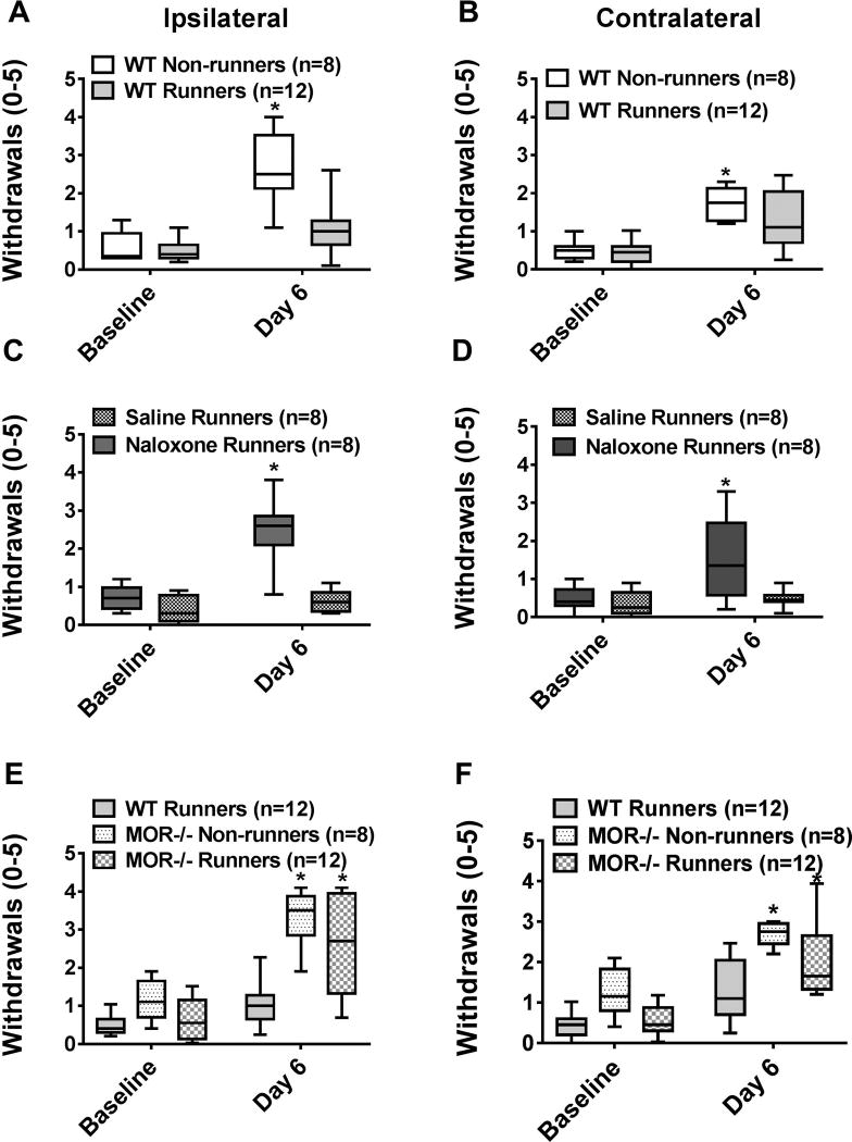
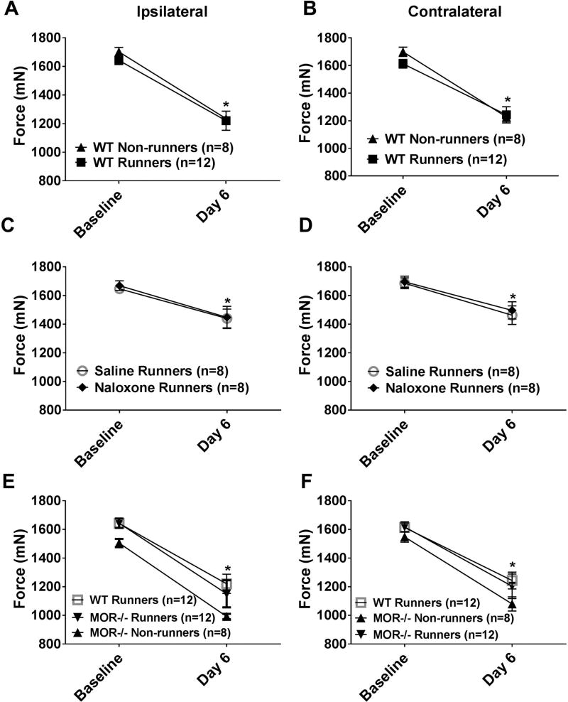
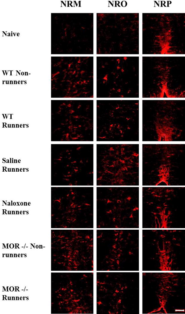
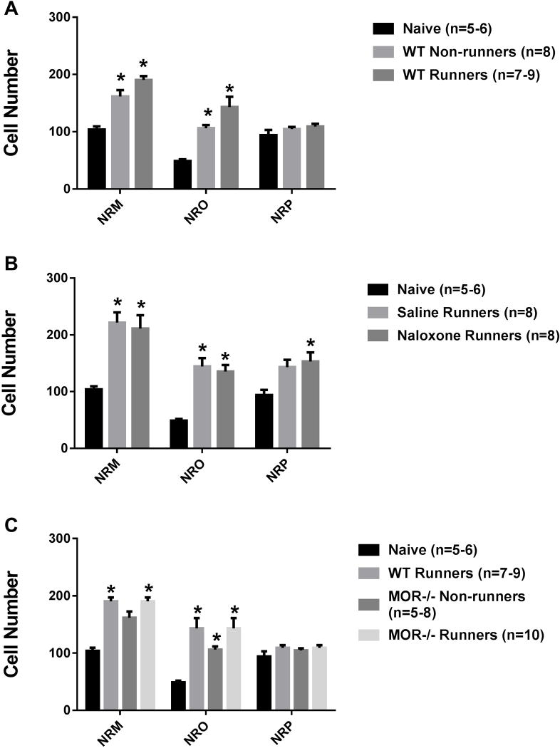
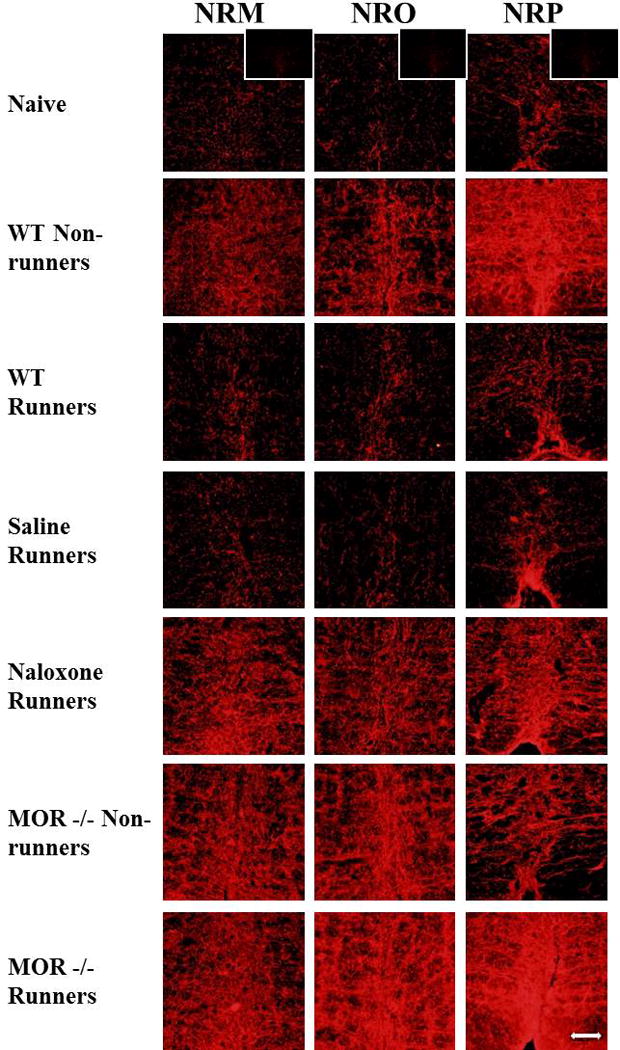
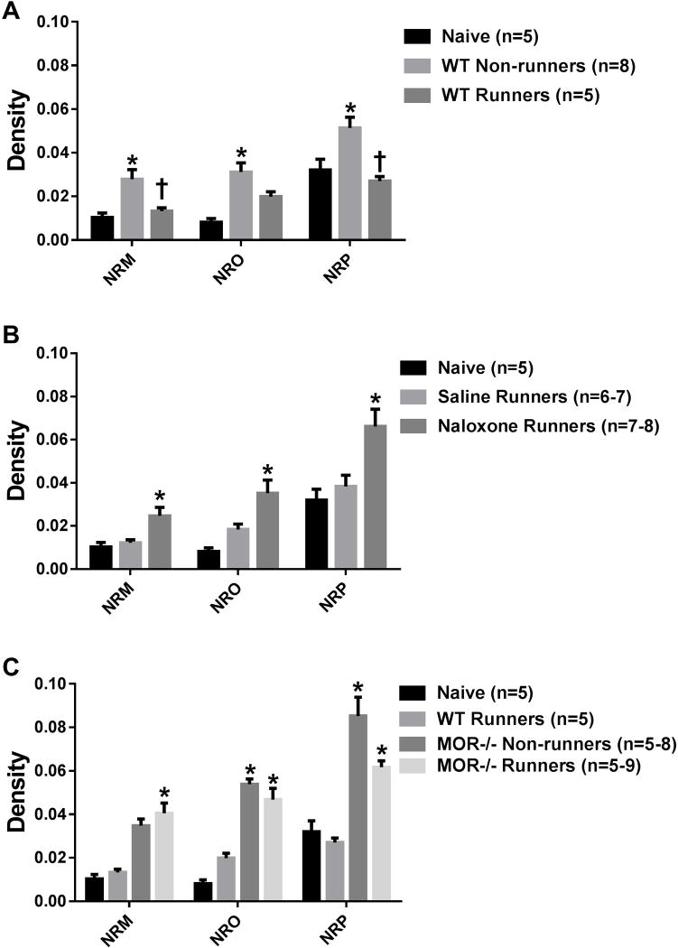
Regular physical activity prevents the development of chronic muscle pain through the modulation of central mechanisms that involve rostral ventromedial medulla (RVM). We tested if pharmacological blockade or genetic deletion of mu-opioid receptors in physically active mice modulates excitatory and inhibitory systems in the RVM in an activity-induced hyperalgesia model. We examined response frequency to mechanical stimulation of the paw, muscle withdrawal thresholds, and expression of phosphorylation of the NR1 subunit of the N-methyl-D-aspartate receptor (p-NR1) and serotonin transporter (SERT) in the RVM. Mice that had performed 5 days of voluntary wheel running prior to the induction of the model were compared with sedentary mice. Sedentary mice showed significant increases in mechanical paw withdrawal frequency and a reduction in muscle withdrawal threshold; wheel running prevented the increase in paw withdrawal frequency. Naloxone-treated and MOR mice had increases in withdrawal frequency that were significantly greater than that in physically active control mice and similar to sedentary mice. Immunohistochemistry in the RVM showed increases in p-NR1 and SERT expression in sedentary mice 24 hours after the induction of the model. Wheel running prevented the increase in SERT, but not p-NR1. Physically active, naloxone-treated, and MOR mice showed significant increases in SERT immunoreactivity when compared with wild-type physically active control mice. Blockade of SERT in the RVM in sedentary mice reversed the activity-induced hyperalgesia of the paw and muscle. These results suggest that analgesia induced by 5 days of wheel running is mediated by mu-opioid receptors through the modulation of SERT, but not p-NR1, in RVM.


Conflict of interest statement

The authors declare no conflict of interest.

Figures

Fig. 1
Fig. 1. Mechanical hyperalgesia of the paw
Graphs represent the number of withdrawals to repeated mechanical stimulation of the hindpaw with an 0.4 mN force for the ipsilateral (A, C, E) and contralateral (B, D, F) sides. A,B. After induction of activity-induced hyperalgesia, the number of responses significantly increases in sedentary WT mice (WT non-runners). 5 days of running wheel activity prevented these increases in the number of responses to mechanical stimuli (WT Runners). *P < 0.05 when compared to WT Non-runners. C,D. The group of mice treated with naloxone during the 5 days of wheel running showed significantly higher responses to mechanical stimulation than saline controls. *P < 0.05 when compared to saline runners. E,F. MOR −/− runners showed significantly greater increases in the number of responses to mechanical stimulation of the paw when compared to WT Runners; responses were similar to MOR−/− non-runners. *P < 0.05 when compared to WT Runners. WT: wild type, MOR−/−: Mu-opioid receptor knockout. Box plots represent the median with the 10th and 90th percentiles.
Fig. 2
Fig. 2. Mechanical hyperalgesia of the muscle
Graphs represent the withdrawal threshold of the muscle for the ipsilateral (A, C, E) and contralateral (B, D, F) sides. A,B. After induction of activity-induced hyperalgesia, the withdrawal threshold of the muscle decreases bilaterally in sedentary WT mice (WT non-runners). 5 days of running wheel activity had no effect on the decreased withdrawal threshold of the muscle (WT Runners). *P < 0.05 when compared to baseline. C,D. Treatment with naloxone during the 5 days of wheel running had no effect on the decreased withdrawal threshold of the muscle when compared to saline treatment. *P < 0.05 when compared to baseline E,F. MOR−/− runners showed significantly greater increases in the number of responses to mechanical stimulation of the paw when compared to WT Runners and MOR−/− non-runners. *P < 0.05 when compared to baseline. WT: wild type, MOR−/−: Mu-opioid receptor knockout. Data are the mean with S.E.M.
Fig. 3
Fig. 3. Photomicrographs of p-NR1 immunohistochemistry
Immunohistochemical staining of p-NR1 in the nucleus raphe magnus (NRM), nucleus raphe obscurus (NRO), and nucleus raphe pallidus (NRP) are represented for each group: Naive, WT-Non-runners, WT Runners, Saline Runners, Naloxone Runners, MOR−/− Non-runners and MOR−/− Runners. Bar represents 50 µm. WT: wild type, MOR−/−: Mu-opioid receptor knockout.
Fig. 4
Fig. 4. P-NR1 staining
Graphs represent the number of immunohistochemically stained p-NR1 in the RVM. A. Increases in the number of p-NR1 positive cells occurred in the NRM and NRO 24h after induction of the pain model in sedentary mice (WT Non-runners). WT Runners showed a similar increase in the number of p-NR1 positive cells after induction of the model. *p < 0.05 when compared to naive. B. Administration of naloxone during the 5 days of running wheel had no effect on the number of p-NR1 positive cells in the RVM. Significant increases in p-NR1 cells occurred in the NRM and the NRO in both the naloxone runners and the saline runners when compared to naive mice (*p < 0.05). C. MOR−/− runner mice had significantly greater number of p-NR1 positive cells when compared to WT runners in the NRM and NRO (*p < 0.05). WT: wild type, MOR−/−: Mu-opioid receptor knockout. Data are the mean with S.E.M.
Fig. 5
Fig. 5. Photomicrographs of immunohistochemistry for the serotonin transporter (SERT)
Immunohistochemical staining for the nucleus raphe magnus (NRM), nucleus raphe obscurus (NRO), and nucleus raphe pallidus (NRP) are represented for each group: Naive, WT-Non-runners, WT Runners, Saline Runners, Naloxone Runners, MOR−/− Non-runners and MOR−/− Runners. Bar represents 50 µm. WT: wild type, MOR−/−: Mu-opioid receptor knockout. *Inset shows photomicrograph from SERT−/− mouse incubated with the antibody to SERT. There was no immunoreactivity for SERT in SERT−/− mice.
Fig. 6
Fig. 6. SERT staining
Graphs represent the quantification of SERT immunoreactivity in the RVM. A. In sedentary mice (WT Non-runners), there was a significant increase in SERT immunoreactivity in the nucleus raphe magnus (NRM), nucleus raphe obscurus (NRO), and nucleus raphe pallidus (NRP) when compared to naive mice (*p < 0.05). This increase did not occur in mice who had done 5 days of wheel running prior to induction of the model in the NRM and the NRP (†p < 0.05). B. Naloxone-treated physically active mice (Naloxone Runners) had a significant increase in SERT immunoreactivity in all three areas when compared to both saline-treated and naive mice (*p < 0.05). C. Physically active MOR−/− mice (MOR−/− Non-runners) had significantly higher SERT immunoreactivity when compared to both physically active WT (WT Runners) and naive mice (*p < 0.05). Sedentary MOR−/− mice had a similar increase in SERT when compared to WT Runners. WT: wild type, MOR−/−: Mu-opioid receptor knockout.
Fig. 7
Fig. 7. Mechanical hyperalgesia of the paw and muscle after microinjection of fluoxetine
Graphs represent the number of withdrawals to repeated mechanical stimulation of the hindpaw with an 0.4 mN force for the ipsilateral (A) and contralateral (B) sides and the withdrawal threshold of the muscle for the ipsilateral (C) and contralateral (D) sides. All groups showed an increase in the paw withdrawal frequency to mechanical stimulation and a decrease in muscle withdrawal thresholds on day 6. Microinjection of fluoxetine (20nmol/0.2ul) into the RVM decreased the paw withdrawal frequency to mechanical stimulation (*p=0.008) and increased the muscle withdrawal threshold (*p=0.005) 15 min after injection when compared to vehicle controls, but not after 2h (p > 0.005). Mice injected with saline in the RVM or fluoxetine outside of the RVM (missed sites) had no reversal of hyperalgesia (p > 0.05). E. Maps showing location of injection sites in the RVM for fluoxetine (red) and saline (blue), and for sites outside the RVM (green) for each individual animal at Bregma levels −5.52 to −6.72.

Similar articles

Cited by

References

    1. Arvidsson U, Cullheim S, Ulfhake B, Ramírez V, Dagerlind Å, Luppi PH, Kitahama K, Jouvet M, Terenius L, Åman K. Distribution of enkephalin and its relation to serotonin in cat and monkey spinal cord and brain stem. Synapse. 1992;11(2):85–104. - PubMed
    1. Baraniuk JN, Whalen G, Cunningham J, Clauw DJ. Cerebrospinal fluid levels of opioid peptides in fibromyalgia and chronic low back pain. BMC Musculoskelet Disord. 2004;5:48. - PMC - PubMed
    1. Basbaum AI, Fields HL. Endogenous pain control systems: brainstem spinal pathways and endorphin circuitry. Annu Rev Neurosci. 1984;7(1):309–338. - PubMed
    1. Bement MKH, Sluka KA. Low-intensity exercise reverses chronic muscle pain in the rat in a naloxone-dependent manner. Arch Phys Med Rehabil. 2005;86(9):1736–1740. - PubMed
    1. Bidonde J, Jean Busch A, Bath B, Milosavljevic S. Exercise for adults with fibromyalgia: an umbrella systematic review with synthesis of best evidence. Current rheumatology reviews. 2014;10(1):45–79. - PubMed

MeSH terms

Substances