. 2017 Aug;23(8):474-484.
doi: 10.1089/ten.TEC.2017.0133.

Human Induced Pluripotent Stem Cell-Derived Endothelial Cells for Three-Dimensional Microphysiological Systems

Affiliations

Human Induced Pluripotent Stem Cell-Derived Endothelial Cells for Three-Dimensional Microphysiological Systems

Yosuke K Kurokawa et al. Tissue Eng Part C Methods. 2017 Aug.

Abstract

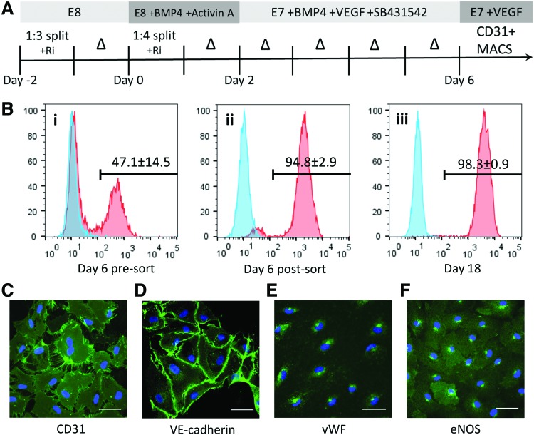
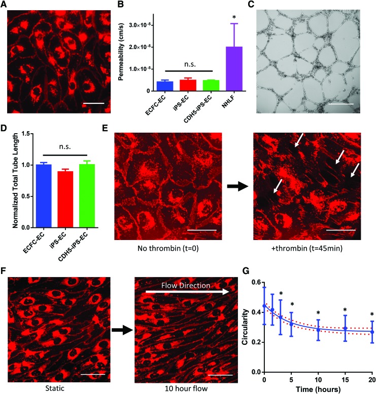
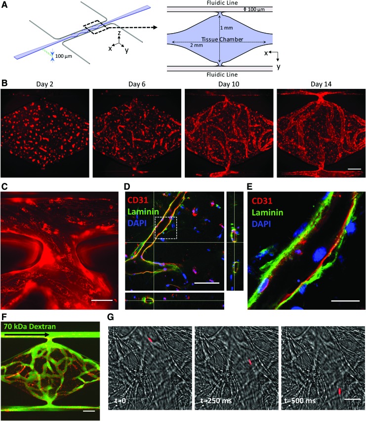
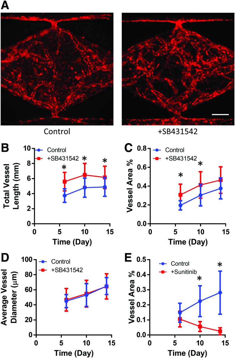
Microphysiological systems (MPS), or "organ-on-a-chip" platforms, aim to recapitulate in vivo physiology using small-scale in vitro tissue models of human physiology. While significant efforts have been made to create vascularized tissues, most reports utilize primary endothelial cells that hinder reproducibility. In this study, we report the use of human induced pluripotent stem cell-derived endothelial cells (iPS-ECs) in developing three-dimensional (3D) microvascular networks. We established a CDH5-mCherry reporter iPS cell line, which expresses the vascular endothelial (VE)-cadherin fused to mCherry. The iPS-ECs demonstrate physiological functions characteristic of primary endothelial cells in a series of in vitro assays, including permeability, response to shear stress, and the expression of endothelial markers (CD31, von Willibrand factor, and endothelial nitric oxide synthase). The iPS-ECs form stable, perfusable microvessels over the course of 14 days when cultured within 3D microfluidic devices. We also demonstrate that inhibition of TGF-β signaling improves vascular network formation by the iPS-ECs. We conclude that iPS-ECs can be a source of endothelial cells in MPS providing opportunities for human disease modeling and improving the reproducibility of 3D vascular networks.

Keywords: endothelial cells; induced pluripotent stem cells; microfluidics; vascularization.


Conflict of interest statement

S.C.G. is a cofounder of 4Design Biosciences, LLC. All other authors have no competing financial interests.

Figures

<b>FIG. 1.</b>
FIG. 1.
Differentiation, isolation, and basic characterization of iPS-ECs. (A) Schematic of the iPS-EC differentiation protocol. The iPS-ECs were isolated using MACS for CD31 on day 6. (B) The differentiated cells were analyzed for CD31 expression by flow cytometry on day 6 (i) before sorting, (ii) after MACS sorting, and (iii) 12 days of culture after sorting. Blue = isotype control. The expression of definitive endothelial markers was confirmed using immunofluorescence for (C) CD31, (D) VE-cadherin, (E) vWF, and (F) eNOS. Scale bar: 50 μm. eNOS, endothelial nitric oxide synthase; iPS-EC, induced pluripotent stem cell-derived endothelial cells; VE, vascular endothelial; vWF, von Willebrand factor. Color images available online at www.liebertpub.com/tec
<b>FIG. 2.</b>
FIG. 2.
Phenotype characterization of iPS-ECs. (A) The ratio of ephrinB2 to EphB4 expression levels measured by qPCR. The expression levels are normalized to ECFC-ECs, with values >0 indicating a more arterial phenotype than ECFC-ECs. HUVECs, HDLECs, and iPS-ECs demonstrate a more venous phenotype than ECFC-ECs. *p < 0.05 compared to iPS-ECs. (B) Expression levels of lymphatic markers measured by qPCR, normalized to ECFC-ECs. *p < 0.05 compared to iPS-ECs. (C) Expression of lymphatic markers confirmed by immunofluorescence. The HDLECs stain positively for all three markers, while the iPS-ECs only stain positively for LYVE-1. Blue = DAPI. Scale bar: 50 μm. DAPI, 4′,6-diamidino-2-phenylindole, dihydrochloride; ECFC-EC, endothelial colony-forming cell-derived endothelial cell; HDLECs, human dermal lymphatic endothelial cells; HUVECs, human umbilical vein endothelial cells; LYVE-1, lymphatic vessel endothelial hyaluronan receptor-1; qPCR, quantitative polymerase chain reaction. Color images available online at www.liebertpub.com/tec
<b>FIG. 3.</b>
FIG. 3.
The use of CDH5-mCherry iPSC-derived ECs (CDH5-iPS-ECs). (A) The CDH5-iPS-ECs demonstrate mCherry signal that localizes to cell junctions. (B) The permeability measured by the diffusion of 70 kDa dextran across a cell monolayer. The permeability of CDH5-iPS-EC monolayer is unchanged compared to iPS-ECs as well as ECFC-ECs. *p < 0.05 compared to CDH5-iPS-ECs. (C) Representative image of Matrigel tube formation assay for CDH5-iPS-ECs. (D) The total tube length normalized to ECFC-ECs. No significant differences were observed across the three EC types. (E) CDH5-iPS-EC response to thrombin. The mCherry-VE-cadherin disaggregate from cell junctions, resulting in the formation of intercellular gaps (indicated by white arrows). (F) The CDH5-iPS-ECs are used to image the response to shear stress in real time. The cells demonstrate a more elongated morphology over time. (G) The circularity of the CDH5-iPS-ECs in response to shear stress is calculated over time. *p < 0.05 compared to time 0. Line indicates a one-phase decay fit with 95% confidence band. Scale bar: 50 μm (A, E, F), 500 μm (C). Color images available online at www.liebertpub.com/tec
<b>FIG. 4.</b>
FIG. 4.
Three-dimensional culture of iPS-ECs in microfluidic devices. (A) Schematic illustration of the microfluidic device. The cell suspension in fibrin is loaded into the tissue chamber region, while media are delivered through the top and bottom fluidic lines. (B) Representative images of the vessel network formation by CDH5-iPS-ECs tracked over a period of 14 days. (C) The vessel anastomoses to the top fluidic line as indicated by the continuous CDH5-iPS-EC lining around the opening of the tissue chamber. (D) The microvessels demonstrate a patent lumen as confirmed by confocal microscopy. The dashed box region: (E) the iPS-ECs deposit laminin as a part of the basement membrane. (F) The vessel network effectively retains 70 kDa dextran introduced through the top fluidic line. (G) One micrometer bead (red) is captured flowing through the vessel. Scale bar: 200 μm (B, F), 50 μm (C), 100 μm (D), 25 μm (E, G). Color images available online at www.liebertpub.com/tec
<b>FIG. 5.</b>
FIG. 5.
Response of iPS-EC vasculature to small molecule inhibitors. (A) Representative CDH5-iPS-EC vessel networks developed at day 14 with or without the supplementation of SB431542 in the growth medium. The addition of SB432542 significantly increased (B) the total vessel length and (C) the total vessel area coverage within the tissue chamber. *p < 0.05 compared to control at the given time point. (D) The average vessel diameter is not significantly affected by the addition of SB431542. (E) The total vessel area within the tissue chamber decreases in response to sunitinib treatment. *p < 0.05 comparing control versus sunitinib-treated condition at the given time point. Color images available online at wwwliebertpub.com/tec

Similar articles

Cited by

References

    1. Cleaver O., and Melton D.A. Endothelial signaling during development. Nat Med 9, 661, 2003 - PubMed
    1. Rafii S., Butler J.M., and Ding B.-S. Angiocrine functions of organ-specific endothelial cells. Nature 529, 316, 2016 - PMC - PubMed
    1. Deanfield J.E., Halcox J.P., and Rabelink T.J. Endothelial function and dysfunction: testing and clinical relevance. Circulation 115, 1285, 2007 - PubMed
    1. Heylman C., Sobrino A., Shirure V.S., Hughes C.C., and George S.C. A strategy for integrating essential three-dimensional microphysiological systems of human organs for realistic anticancer drug screening. Exp Biol Med 239, 1240, 2014 - PMC - PubMed
    1. Esch M.B., Smith A.S.T., Prot J.-M., Oleaga C., Hickman J.J., and Shuler M.L. How multi-organ microdevices can help foster drug development. Adv Drug Deliv Rev 69–70, 158, 2014 - PMC - PubMed

Publication types

MeSH terms