Review
. 2017 Sep;24(9):1478-1487.
doi: 10.1038/cdd.2017.82. Epub 2017 Jun 16.

BCL-2 family: integrating stress responses at the ER to control cell demise

Affiliations
Review

BCL-2 family: integrating stress responses at the ER to control cell demise

Philippe Pihán et al. Cell Death Differ. 2017 Sep.

Abstract

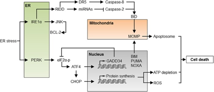
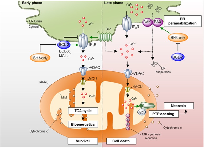
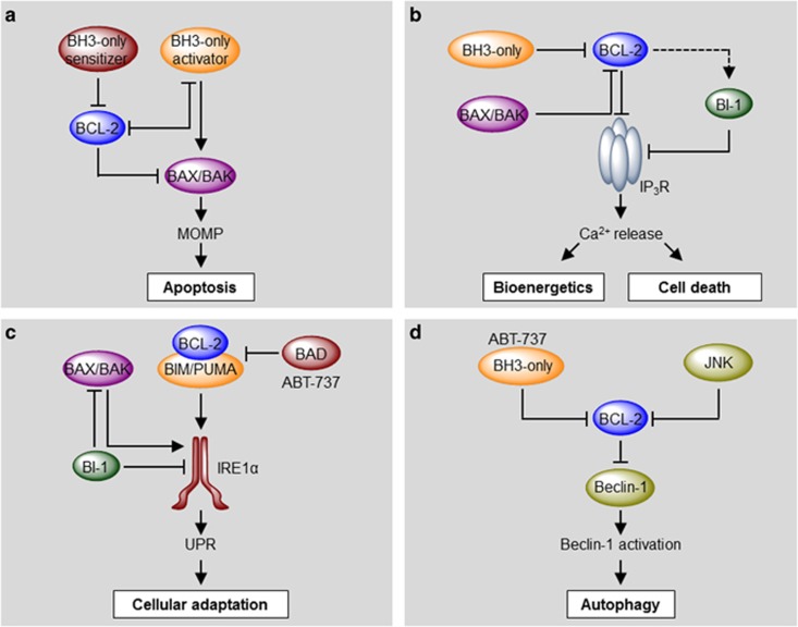
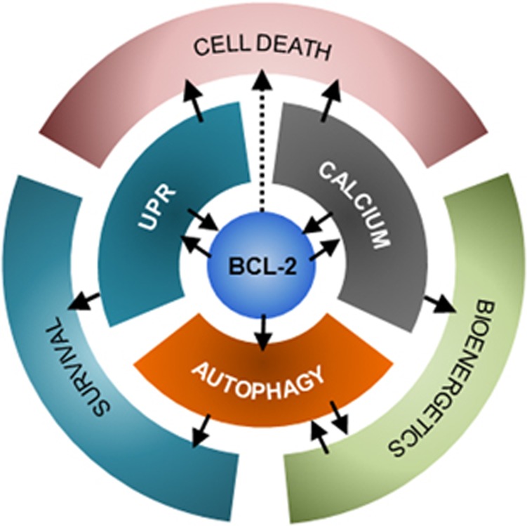
In the last decade, the endoplasmic reticulum (ER) has emerged as a central organelle regulating the core mitochondrial apoptosis pathway. At the ER membrane, a variety of stress signals are integrated toward determining cell fate, involving a complex cross talk between key homeostatic pathways including the unfolded protein response, autophagy, calcium signaling and mitochondrial bioenergetics. In this context, key regulators of cell death of the BCL-2 and TMBIM/BI-1 family of proteins have relevant functions as stress rheostats mediated by the formation of distinct protein complexes that regulate the switch between adaptive and proapoptotic phases under stress. Here, we overview recent advances on our molecular understanding of how the apoptotic machinery integrates stress signals toward cell fate decisions upstream of the mitochondrial gateway of death.


Conflict of interest statement

The authors declare no conflict of interest.

Figures

Figure 1
Figure 1
Regulation of apoptosis by the BCL-2 family. (a) The BCL-2 family of proteins is divided into three groups based on their functional role in the regulation of apoptosis and the number of BCL-2 homology (BH) domains they bear. (b) The BCL-2 proteins regulate the permeabilization of the mitochondrial outer membrane (MOMP) and apoptosis through a specific network of heterodimeric interactions. In one model, termed the direct activation model, a subset of BH3-only proteins directly engages and activates BAX or BAK leading to MOMP. Prosurvival BCL-2 proteins suppress apoptosis through their interaction with and sequestration of these ‘activator’ BH3-only proteins. Other BH3-only proteins – denominated ‘sensitizers’ – may competitively interact with prosurvival proteins, releasing activator BH3-only proteins. In the neutralization model, constitutively active BAX and BAK are kept in check by prosurvival BCL-2 proteins. Upon activation, BH3-only proteins interact with and ‘neutralize’ prosurvival BCL-2 proteins, releasing BAX and BAK and engaging MOMP. It is likely that aspect of both models contribute to the regulation of apoptosis under physiological conditions. BAD, BCL-2 antagonist of cell death; BAK, BCL-2 antagonist or killer; BAX, BCL-2-associated X protein; BID, BH3-interacting domain death agonist; BIK, BCL-2-interacting killer; BIM, BCL-2-interacting mediator of cell death; BMF, BCL-2-modifying factor; BNIP3, BCL-2 and adenovirus E1B 19 kDa protein-interacting protein 3; HRK, harakiri; PUMA, p53-upregulated modulator of apoptosis; TM, transmembrane. *NOXA and BMF have also been shown to exhibit direct activation function under certain circumstances
Figure 2
Figure 2
The apoptosis regulatory network under ER stress. Under acute or sustained ER stress, the UPR actively promotes apoptosis through the regulation of proapoptotic proteins of the BCL-2 family, increased proteotoxicity and ROS. IRE1α activation engages the degradation of several ER-localized RNAs through RIDD, having a dual role in apoptosis regulation. Degradation of several inhibitory miRNAs leads to the activation of caspase-2, which in turn cleaves and activates the BH3-only protein BID triggering the mitochondrial pathway of apoptosis. Conversely, degradation of the DR5 mRNA results in the inhibition of caspase-8, repressing cell death. JNK is also activated downstream of IRE1α, inducing cell death through BCL-2 phosphorylation and inhibition. Activation of PERK leads to the phosphorylation of eIF2α and global protein synthesis arrest. Under these conditions, ATF4 is selectively translated and, together with CHOP, transcriptionally activates several BH3-only proteins, engaging the mitochondria. Moreover, the ATF4/CHOP complex resumes protein synthesis through the upregulation of the GADD34 phosphatase and other genes, leading to ATP depletion, ROS and finally cell death. ATF4, activating transcription factor 4; DR5, death receptor 5; GADD34, growth arrest and DNA damage-inducible protein 34; JNK, c-Jun N-terminal kinase; RIDD, regulated IRE1-dependent decay
Figure 3
Figure 3
Regulation of ER calcium homeostasis by the BCL-2 family. Different pro- and anti-apoptotic proteins of the BCL-2 family localize to the ER membrane where they regulate ER calcium homeostasis mainly through the modulation of the IP3Rs. Left panel: under physiological conditions, anti-apoptotic proteins of the BCL-2 family interact with the IP3R decreasing its calcium leak activity or promoting the release of oscillatory calcium bursts. This calcium signaling is transduced to the mitochondrial matrix through the VDAC and MCU1 channels at the mitochondria outer and inner membranes, respectively, where it promotes the activity of several metabolic enzymes, increasing ATP production and mitochondrial bioenergetics. The regulation of the IP3R by BCL-2 proteins may be antagonized by BH3-only proteins and BAX. BI-1, a protein of the TMBIM family, also interacts with IP3R, diminishing ER-to-mitochondria calcium transfer. Right panel: under some apoptotic conditions, IP3Rs are activated, leading to sustained calcium release from the ER. Massive calcium uptake by the mitochondria leads to the opening of the mPTP, resulting in mitochondrial swelling, MOMP and decrease in ATP production. This leads to necrotic cell death. Additionally, the ER membrane becomes permeabilized, leading to the release of calcium- and ER-resident proteins. This process is regulated by the proteins of the BCL-2 family. IP3R, inositol 1,4,5-trisphosphate receptor; MCU, mitochondrial calcium uniporter; VDAC, voltage-dependent anion channel
Figure 4
Figure 4
Molecular hierarchical regulation of apoptosis, calcium, the UPR and autophagy by the BCL-2 family of proteins. Different pro- and anti-apoptotic members of the BCL-2 family regulate apoptosis, ER calcium homeostasis, the UPR and autophagy by a complex network of protein–protein interactions with different functional hierarchical organizations. (a) In a combined model of apoptosis regulation by the BCL-2 family, BH3-only activators may directly engage BAX or BAK, causing their homo-oligomerization and MOMP. Anti-apoptotic proteins of the BCL-2 family bind to and sequester activator BH3-only proteins, inhibiting cell death. This inhibitory function may be reversed by sensitizer BH3-only proteins. (b) At the ER membrane, anti-apoptotic BCL-2 interacts with IP3R inhibiting its calcium leak channel activity, leading to low transient calcium release and improved mitochondrial bioenergetics. This inhibition may be antagonized by BAX, BAK and proapoptotic BH3-only proteins, increasing ER-to-mitochondrial calcium transfer and triggering mPTP-driven necrosis/apoptosis. Additionally, BI-1 also interacts with and blocks IP3R, probably downstream of BCL-2 proteins. (c) BAX, BAK, BCL-2 and BH3-only proteins BIM and PUMA activate IRE1α signaling upon ER stress. Interestingly, this function depends on the formation of a protein complex between BIM/PUMA and BCL-2, and can be inhibited by BAD or the BH3 mimetics ABT-737. BI-1 inhibits IRE1α and BAX/BAK. (d) Anti-apoptotic proteins of the BCL-2 family inhibit autophagy through the interaction with Beclin-1. BH3-only proteins or ABT-737 may displace this inhibitory interaction, releasing Beclin-1 and inducing autophagy. Interestingly, the IRE1α/JNK pathway may also induce autophagy through the phosphorylation and inhibition of BCL-2
Figure 5
Figure 5
Integrating stress responses at the ER. The ER pool of proteins of the BCL-2 family plays a central role in the regulation of the UPR, calcium homeostasis and autophagy, acting as molecular bridges between these stress pathways. Additionally, the activation of these pathways may lead to different cellular outcomes, such as improved bioenergetics and survival or cell death. Bidirectional regulations and cross talk are indicated with arrows. Thus, the BCL-2 family at the ER plays a pivotal role in cell fate determination upon ER stress

References

    1. Youle RJ, Strasser A. The BCL-2 protein family: opposing activities that mediate cell death. Nat Rev Mol Cell Biol 2008; 9: 47–59. - PubMed
    1. Tait SW, Green DR. Mitochondria and cell death: outer membrane permeabilization and beyond. Nat Rev Mol Cell Biol 2010; 11: 621–632. - PubMed
    1. Kerr JF, Wyllie AH, Currie AR. Apoptosis: a basic biological phenomenon with wide-ranging implications in tissue kinetics. Br J Cancer 1972; 26: 239–257. - PMC - PubMed
    1. Taylor RC, Cullen SP, Martin SJ. Apoptosis: controlled demolition at the cellular level. Nat Rev Mol Cell Biol 2008; 9: 231–241. - PubMed
    1. Galluzzi L, Bravo-San Pedro JM, Kroemer G. Organelle-specific initiation of cell death. Nat Cell Biol 2014; 16: 728–736. - PubMed

MeSH terms

Substances

LinkOut - more resources