Review
. 2017 Jul;23(7):615-635.
doi: 10.1016/j.molmed.2017.05.006. Epub 2017 Jun 13.

Pathogenesis of Human Systemic Lupus Erythematosus: A Cellular Perspective

Affiliations
Review

Pathogenesis of Human Systemic Lupus Erythematosus: A Cellular Perspective

Vaishali R Moulton et al. Trends Mol Med. 2017 Jul.

Abstract

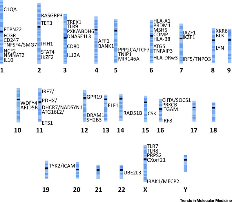
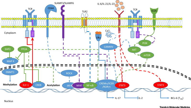
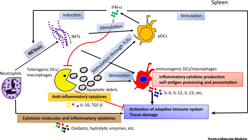
Systemic lupus erythematosus (SLE) is a chronic autoimmune disease affecting multiple organs. A complex interaction of genetics, environment, and hormones leads to immune dysregulation and breakdown of tolerance to self-antigens, resulting in autoantibody production, inflammation, and destruction of end-organs. Emerging evidence on the role of these factors has increased our knowledge of this complex disease, guiding therapeutic strategies and identifying putative biomarkers. Recent findings include the characterization of genetic/epigenetic factors linked to SLE, as well as cellular effectors. Novel observations have provided an improved understanding of the contribution of tissue-specific factors and associated damage, T and B lymphocytes, as well as innate immune cell subsets and their corresponding abnormalities. The intricate web of involved factors and pathways dictates the adoption of tailored therapeutic approaches to conquer this disease.

Keywords: SLE; autoimmunity; immune cells.


Figures

Figure 1
Figure 1
Genetics of Systemic Lupus Erythematosus (SLE). The schematic depicts a panel of chromosomes showing genes associated with SLE. The approximate positions of SLE-associated loci and genes in the human genome are shown. These genes were selected because they have been validated in two or more studies. FCGR includes FCGR2A, FCGR3B, and FCGR3A; COMP includes C2, C4A, and C4B.
Figure 2
Figure 2
T Cell Signaling and Gene Regulation Defects in Systemic Lupus Erythematosus (SLE). In SLE patients, increased protein phosphatase 2A (PP2A) levels and activity in T cells suppresses Ets-like-factor-1 (ELF-1), a transcriptional enhancer of the CD3ζ chain and a repressor of the FcεRIγ (Fcγ) chain. TCR/CD3 complex rewiring, via replacement of CD3ζ with Fcγ, results in increased calcium responses, enhancing the activity of calcium/calmodulin-dependent protein kinase IV (CaMKIV), which in turn increases the binding of cAMP response element modulator (CREMα) and inducible cAMP early repressor (ICER). CREMα is also activated by ERα, and is recruited to the IL17A and IL2 promoters, enhancing and repressing their transcription, respectively [185,194]. It is known that CaMIV activity is enhanced by the presence of costimulatory molecules such as ICOS in preformed lipid-rafts containing the TCR, which can lead to activation of the PI3K/mTOR pathway; enhanced activity of protein phosphatase 2A (PP2A) and SHP2 suppresses the MAPK–DNMT1 pathway and dephosphorylates cAMP-responsive element-binding protein 1 (CREB), resulting in suppression of IL2 transcription [189]. PP2A also acts through Rho-associated protein kinase (ROCK) to enhance binding of the IL17 transcription enhancer interferon regulatory factor 4 (IRF4); signaling through CD44 also activates ROCK, which promotes cell migration and binding of IRF4 to the IL17 promoter [49,51]. TLRs and SLAMF signaling can activate the transcription factors NF-κB and NFAT, and this contributes to the transcription of proinflammatory cytokines [201]. In addition, pro-inflammatory cytokines IL-6, IL-21, and IL-23 cause downstream activation of STAT3 transcriptional targets, including IL17 and BCL6, contributing to inflammation and supporting antibody production from B cells Abbreviations: [46,52]. ER, estrogen receptor; ICOS, inducible T cell costimulator; SHP2, Src homology 2 domain containing phosphotyrosine phosphatase 2; TCR, T cell receptor; Tfh, T follicular helper cells; TLR, Toll-like receptor.
Figure 3
Figure 3
Role of Innate Cells (Neutrophils, Macrophages and Dendritic Cells) in Systemic Lupus Erythematosus (SLE) Pathogenesis. Altered functional properties in neutrophils have been observed in SLE, including decreased clearance of apoptotic material and increased synthesis and release of various proteins including oxidants, hydrolytic enzymes, and inflammatory cytokines which contribute to tissue damage. Myeloid dendritic cells (DCs) and plasmacytoid dendritic cells (pDCs) represent two major DC subsets derived from different developmental pathways from their precursors. pDCs are a specialized type of interferon (IFN)-producing cells capable of producing massive amounts of type-I IFNs upon stimulation. Type I IFNs can induce the formation of neutrophil extracellular traps (NETs), which are a source of self-stimuli and reciprocally enhance production of type I IFNs [104]. Based on their function, myeloid DCs can be further divided into tolerogenic and immunogenic DCs. In SLE patients, immunogenic DCs/macrophages acquire activated phenotypes with increased production of inflammatory cytokines or enhanced self-antigen processing and presentation. Tolerogenic DC/macrophages are responsible for the rapid removal of apoptotic cells, and, coupled with their ability to produce anti-inflammatory cytokines, efficiently suppress autoimmunity. In SLE patients, a significant reduction in both the number and function of these cells is observed [105]. Abbreviations: BDCA2–DTR, blood dendritic cell antigen 2–diphtheria toxin receptor transgene; CAMKIV, calcium/calmodulin-dependent protein kinase type IV; DNMT, DNA methyl transferase; HDAC, histone deacetylase; ICOS, inducible T cell costimulatory; IRF, interferon regulatory factor; PD-1, programmed death receptor; LAP, microtubule-associated protein 1A/1B-light chain 3 (LC3)-associated phagocytosis; mTOR, mechanistic target of rapamycin; NET, neutrophil extracellular trap; PP2A, protein phosphatase 2A; PTPN22, protein tyrosine phosphatase, non-receptor type 22; ROCK, Rho-associated protein kinase; SLAM, signaling lymphocyte activation molecule family of receptors; STAT, signal transducer and activator of transcription; Tfh, T follicular helper cell; TLR, Toll-like receptor.

References

    1. Tsokos GC. Systemic lupus erythematosus. N. Engl. J. Med. 2011;365:2110–2121. - PubMed
    1. Tan EM, et al. The 1982 revised criteria for the classification of systemic lupus erythematosus. Arthritis Rheum. 1982;25:1271–1277. - PubMed
    1. Teruel M, Alarcon-Riquelme ME. The genetic basis of systemic lupus erythematosus: what are the risk factors and what have we learned. J. Autoimmun. 2016;74:161–175. - PubMed
    1. Moulton VR, Tsokos GC. T cell signalling abnormalities contribute to aberrant immune cell function and auto-immunity. J. Clin. Invest. 2015;125:2220–2227. - PMC - PubMed
    1. Tsokos GC, et al. New insights into the immunopatho-genesis of systemic lupus erythematosus. Nat. Rev. 2016;12:716–730. - PubMed

Publication types

MeSH terms