. 2017 Jun 16;7(1):3751.
doi: 10.1038/s41598-017-03864-w.

Epigenetic and antitumor effects of platinum(IV)-octanoato conjugates

Affiliations

Epigenetic and antitumor effects of platinum(IV)-octanoato conjugates

Vojtech Novohradsky et al. Sci Rep. .

Abstract

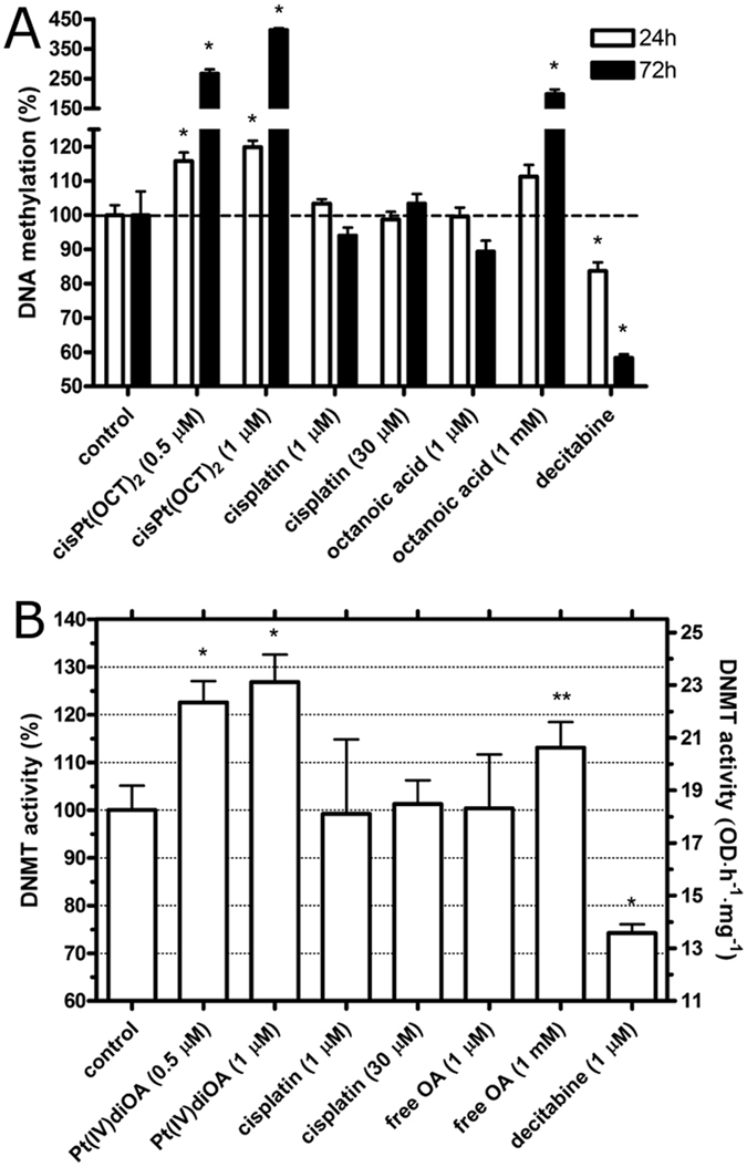
We present the anticancer properties of cis, cis, trans-[Pt(IV)(NH3)2Cl2(OA)2] [Pt(IV)diOA] (OA = octanoato), Pt(IV) derivative of cisplatin containing two OA units appended to the axial positions of a six-coordinate Pt(IV) center. Our results demonstrate that Pt(IV)diOA is a potent cytotoxic agent against many cancer cell lines (the IC50 values are approximately two orders of magnitude lower than those of clinically used cisplatin or Pt(IV) derivatives with biologically inactive axial ligands). Importantly, Pt(IV)diOA overcomes resistance to cisplatin, is significantly more potent than its branched Pt(IV) valproato isomer and exhibits promising in vivo antitumor activity. The potency of Pt(IV)diOA is a consequence of several factors including enhanced cellular accumulation correlating with enhanced DNA platination and cytotoxicity. Pt(IV)diOA induces DNA hypermethylation and reduces mitochondrial membrane potential in cancer cells at levels markedly lower than the IC50 value of free OA suggesting the synergistic action of platinum and OA moieties. Collectively, the remarkable antitumor effects of Pt(IV)diOA are a consequence of the enhanced cellular uptake which makes it possible to simultaneously accumulate high levels of both cisplatin and OA in cells. The simultaneous dual action of cisplatin and OA by different mechanisms in tumor cells may result in a markedly enhanced and unique antitumor effects of Pt(IV) prodrugs.


Conflict of interest statement

The authors declare that they have no competing interests.

Figures

Figure 1
Figure 1
Schematic representation of the platinum compounds used in the present work.
Figure 2
Figure 2
Increase of cytotoxicity, intracellular accumulation and DNA platination in A2780 (A,C) and HCT-116 (B,D) cells treated with Pt(IV)diOA in comparison with the treatment with cisplatin (A,B) or Pt(IV)diAc (C,D).
Figure 3
Figure 3
Activity of HDAC enzymes in HCT-116 cells treated with the compounds tested in the present work; their concentration was 1 µM except the concentration of free OA, which was 1 mM. Symbols: ■, control; ▲, Pt(IV)diOA; ▼, Pt(IV)AcOA; ⧫, Pt(IV)diAc; ●, cisplatin; □, OA (1 mM); ▽, trichostatin A (well known HDAC inhibitor used as the positive control). The data represent a mean from three independent experiments, each performed in quadruplicate. The error bars are the SDs.
Figure 4
Figure 4
Analysis of DNA methylation. (A) DNA was isolated from HCT-116 cells after the exposure to the agents at the concentrations indicated in the graph for 24 or 72 h. The levels of DNA methylation in the control was taken as 100%. The bars represent mean values from three independent experiments each performed in octuplicate. The error bars are the SDs. (B) DNMT activity in colon carcinoma cells HCT-116 treated for 24 h with the compounds tested in the present work. The DNMT activity in the control, untreated cells was taken as 100%. Error bars are the SDs and the stars at the top of bars denote a significant difference from the control with (*p < 0.05; **p < 0.1), calculated using nonparametric student’s t-test. The bars represent mean values from three independent experiments, each performed in quadruplicate.
Figure 5
Figure 5
Confocal microphotographs of HCT-116 cells with nucleus stained with Hoechst 33342 (A) and mitochondria stained with TMRE (B). Cells were treated for 5 h with 1 µM of Pt(IV)diOA (upper row I). Control, untreated cells are shown in the row II (bottom) of the figure. Cells stained with Hoechst 33342 or TMRE were visualized in the bright field mode (C). Scale bars represent 20 µm. Figures are the representatives of at least 10 individual scans of different sample areas. Three independent experiments were performed in triplicate.

References

    1. Hall MD, Hambley TW. Platinum(IV) antitumour compounds: their bioinorganic chemistry. Coord. Chem. Rev. 2002;232:49–67. doi: 10.1016/S0010-8545(02)00026-7. - DOI
    1. Hall MD, Mellor HR, Callaghan R, Hambley TW. Basis for design and development of platinum(IV) anticancer complexes. J. Med. Chem. 2007;50:3403–3411. doi: 10.1021/jm070280u. - DOI - PubMed
    1. Nemirovski A, Kasherman Y, Tzaraf Y, Gibson D. Reduction of cis, trans, cis-PtCl2(OCOCH3)2(NH3)2 by aqueous extracts of cancer cells. J. Med. Chem. 2007;50:5554–5556. doi: 10.1021/jm070740j. - DOI - PubMed
    1. Zhang JZ, et al. Getting to the core of platinum drug bio-distributions: the penetration of anti-cancer platinum complexes into spheroid tumour models. Metallomics. 2012;4:1209–1217. doi: 10.1039/c2mt20168b. - DOI - PubMed
    1. Ravera M, et al. Cellular trafficking, accumulation and DNA platination of a series of cisplatin-based dicarboxylato Pt(IV) prodrugs. J. Inorg. Biochem. 2015;150:1–8. doi: 10.1016/j.jinorgbio.2015.05.012. - DOI - PubMed

Publication types

MeSH terms