

A validated clinical MRI injury scoring system in neonatal hypoxic-ischemic encephalopathy
- PMID: 28623417
- PMCID: PMC6219383
- DOI: 10.1007/s00247-017-3893-y
A validated clinical MRI injury scoring system in neonatal hypoxic-ischemic encephalopathy
Abstract
Background: Deep nuclear gray matter injury in neonatal hypoxic-ischemic encephalopathy (HIE) is associated with worse neurodevelopmental outcomes. We previously published a qualitative MRI injury scoring system utilizing serial T1-weighted, T2-weighted and diffusion-weighted imaging (DWI), weighted for deep nuclear gray matter injury.
Objectives: To establish the validity of the MRI scoring system with neurodevelopmental outcome at 18-24 months.
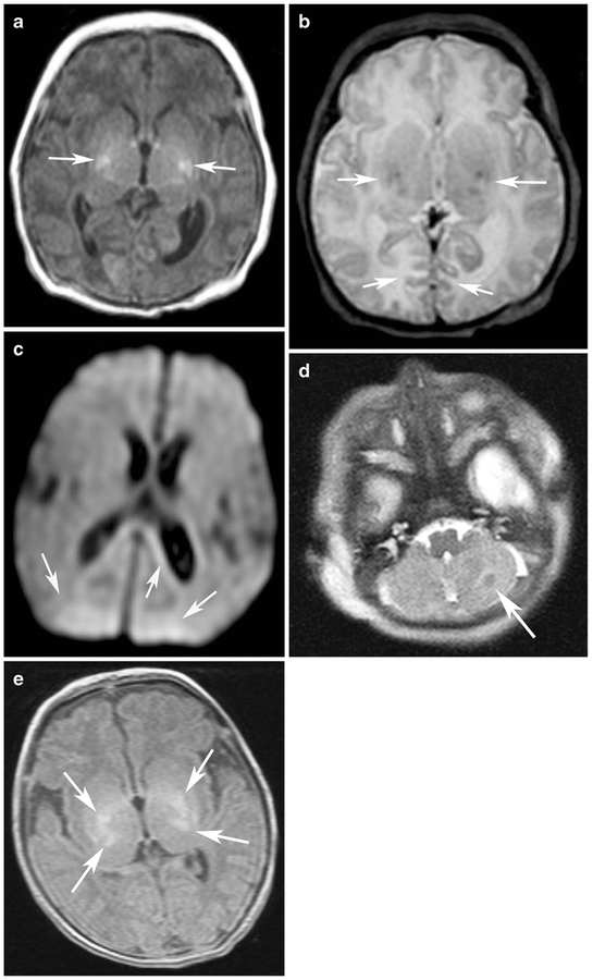
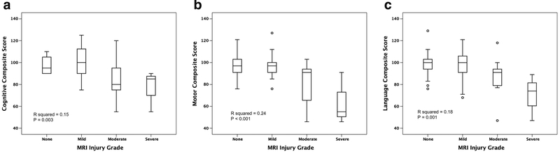
Materials and methods: MRI scans from neonates with moderate to severe HIE treated with therapeutic hypothermia were evaluated. Signal abnormality was scored on T1-weighted, T2-weighted and DWI sequences and assessed using an established system in five regions: (a) subcortical: caudate nucleus, globus pallidus and putamen, thalamus and the posterior limb of the internal capsule; (b) white matter; (c) cortex, (d) cerebellum and (e) brainstem. MRI injury was graded as none, mild, moderate or severe. Inter-rater reliability was tested on a subset of scans by two independent and blinded neuroradiologists. Surviving infants underwent the Bayley Scales of Infant and Toddler Development-III (Bayley-III) at 18-24 months. Data were analyzed using univariate and multivariate linear and logistic regression.
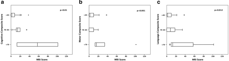
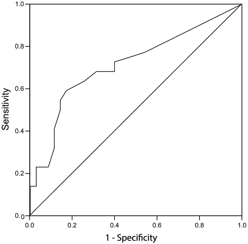
Results: Fifty-seven eligible neonates underwent at least one MRI scan in the first 2 weeks of life. Mean postnatal age at scan 1 was 4±2 days in 50/57 (88%) neonates and 48/54 (89%) surviving infants underwent scan 2 at 10±2 days. In 54/57 (95%) survivors, higher MRI injury grades were significantly associated with worse outcomes in the cognitive, motor and language domains of the Bayley-III.
Conclusion: A qualitative MRI injury scoring system weighted for deep nuclear gray matter injury is a significant predictor of neurodevelopmental outcome at 18-24 months in neonates with HIE.
Keywords: Brain; Hypoxic-ischemic encephalopathy; Magnetic resonance imaging; Neonates; Neurodevelopmental outcome; Scoring system.
Conflict of interest statement
Compliance with ethical standards
Figures
References
-
- Chau V, Poskitt KJ, Dunham CP et al. (2014) Magnetic resonance imaging in the encephalopathic term newborn. Curr Pediatr Rev 10: 28–36 - PubMed
-
- Volpe J (2001) Hypoxic-ischemic encephalopathy: clinical aspects in neurology of the newborn. WBS Company, Philadelphia
-
- Cowan F, Rutherford M, Groenendaal F et al. (2003) Origin and timing of brain lesions in term infants with neonatal encephalopathy. Lancet 361:736–742 - PubMed
-
- Miller SP, Ramaswamy V, Michelson D et al. (2005) Patterns of brain injury in term neonatal encephalopathy. J Pediatr 146:453–460 - PubMed
MeSH terms
Grants and funding
LinkOut - more resources
Full Text Sources
Other Literature Sources
Medical
