Review
doi: 10.17305/bjbms.2017.2083.
Craniosynostosis - Recognition, clinical characteristics, and treatment
Affiliations
- PMID: 28623672
- PMCID: PMC5988529
- DOI: 10.17305/bjbms.2017.2083
Item in Clipboard
Review
Craniosynostosis - Recognition, clinical characteristics, and treatment
Bosn J Basic Med Sci.
.
Abstract
Craniosynostosis is a developmental craniofacial anomaly, resulting in impairment of brain development and abnormally shaped skull. The main cause of craniosynostosis is premature closure of one or more cranial sutures. It usually occurs as an isolated condition, but may also be associated with other malformations as part of complex syndromes. When left untreated, craniosynostosis can cause serious complications, such as developmental delay, facial abnormality, sensory, respiratory and neurological dysfunction, anomalies affecting the eye, and psychological disturbances. Thus, early diagnosis, expert surgical techniques, postoperative care, and adequate follow-up are of vital importance in treating craniosynostosis.
Figures
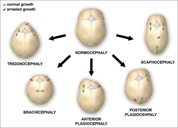
Various deformations of the skull, associated with single-suture synostoses
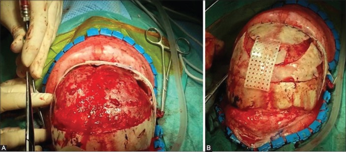
The surgical reconstruction of anterior plagiocephaly. During the operation, the child is placed in a supine position. A coronal skin incision is performed and after the periosteal dissection, the fused right coronal suture is recognized. The bone flap is removed and an extensive remodeling of the orbital bone and forehead follows (A). The final appearance of the skull after the completed surgical reconstruction, filling of bone defects, and their connection with resorbable plates (B).
References
-
- Kjaer I. Neuro-osteology. Crit Rev Oral Biol Med. 1998;9(2):224–44. https://doi.org/10.1177/10454411980090020501. - PubMed
-
- Chai Y, Maxson RE., Jr Recent advances in craniofacial morphogenesis. Dev Dyn. 2006;235(9):2353–75. https://doi.org/10.1002/dvdy.20833. - PubMed
-
- Nah HD, Pacifici M, Gerstenfeld LC, Adams SL, Kirsch T. Transient chondrogenic phase in the intramembranous pathway during normal skeletal development. J Bone Miner Res. 2000;15(3):522–33. https://doi.org/10.1359/jbmr.2000.15.3.522. - PubMed
-
- Gong SG. Cranial neural crest: Migratory cell behaviour and regulatory networks. Exp Cell Res. 2014;325(2):90–5. https://doi.org/10.1016/j.yexcr.2014.03.015. - PubMed
-
- Kouskoura T, Fragou N, Alexiou M, John N, Sommer L, Graf D, et al. The genetic basis of craniofacial and dental abnormalities. Schweiz Monatsschr Zahnmed. 2011;121(7-8):636–46. - PubMed
Publication types
MeSH terms
LinkOut - more resources
Full Text Sources
Other Literature Sources