Characterization of MicroRNA-200 pathway in ovarian cancer and serous intraepithelial carcinoma of fallopian tube
- PMID: 28623900
- PMCID: PMC5473983
- DOI: 10.1186/s12885-017-3417-z
Characterization of MicroRNA-200 pathway in ovarian cancer and serous intraepithelial carcinoma of fallopian tube
Abstract
Background: Ovarian cancer is the leading cause of death among gynecologic diseases in Western countries. We have previously identified a miR-200-E-cadherin axis that plays an important role in ovarian inclusion cyst formation and tumor invasion. The purpose of this study was to determine if the miR-200 pathway is involved in the early stages of ovarian cancer pathogenesis by studying the expression levels of the pathway components in a panel of clinical ovarian tissues, and fallopian tube tissues harboring serous tubal intraepithelial carcinomas (STICs), a suggested precursor lesion for high-grade serous tumors.
Methods: RNA prepared from ovarian and fallopian tube epithelial and stromal fibroblasts was subjected to quantitative real-time reverse-transcription polymerase chain reaction (qRT-PCR) to determine the expression of miR-200 families, target and effector genes and analyzed for clinical association. The effects of exogenous miR-200 on marker expression in normal cells were determined by qRT-PCR and fluorescence imaging after transfection of miR-200 precursors.
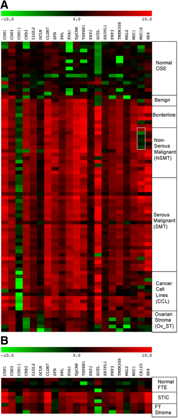
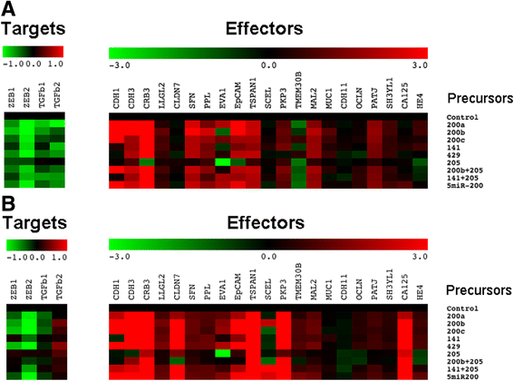
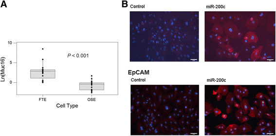
Results: Ovarian epithelial tumor cells showed concurrent up-regulation of miR-200, down-regulation of the four target genes (ZEB1, ZEB2, TGFβ1 and TGFβ2), and up-regulation of effector genes that were negatively regulated by the target genes. STIC tumor cells showed a similar trend of expression patterns, although the effects did not reach significance because of small sample sizes. Transfection of synthetic miR-200 precursors into normal ovarian surface epithelial (OSE) and fallopian tube epithelial (FTE) cells confirmed reduced expression of the target genes and elevated levels of the effector genes CDH1, CRB3 and EpCAM in both normal OSE and FTE cells. However, only FTE cells had a specific induction of CA125 after miR-200 precursor transfection.
Conclusions: The activation of the miR-200 pathway may be an early event that renders the OSE and FTE cells more susceptible to oncogenic mutations and histologic differentiation. As high-grade serous ovarian carcinomas (HGSOC) usually express high levels of CA125, the induction of CA125 expression in FTE cells by miR-200 precursor transfection is consistent with the notion that HGSOC has an origin in the distal fallopian tube.
Keywords: Expression analysis; Fallopian tube tumors; MicroRNA; Ovarian tumors.
Figures


Similar articles
-
Are all pelvic (nonuterine) serous carcinomas of tubal origin?Am J Surg Pathol. 2010 Oct;34(10):1407-16. doi: 10.1097/PAS.0b013e3181ef7b16. Am J Surg Pathol. 2010. PMID: 20861711
-
Both fallopian tube and ovarian surface epithelium are cells-of-origin for high-grade serous ovarian carcinoma.Nat Commun. 2019 Nov 26;10(1):5367. doi: 10.1038/s41467-019-13116-2. Nat Commun. 2019. PMID: 31772167 Free PMC article.
-
Gene expression profiles of ovarian low-grade serous carcinoma resemble those of fallopian tube epithelium.Gynecol Oncol. 2017 Dec;147(3):634-641. doi: 10.1016/j.ygyno.2017.09.029. Epub 2017 Sep 29. Gynecol Oncol. 2017. PMID: 28965696
-
The fallopian tube, "precursor escape" and narrowing the knowledge gap to the origins of high-grade serous carcinoma.Gynecol Oncol. 2019 Feb;152(2):426-433. doi: 10.1016/j.ygyno.2018.11.033. Epub 2018 Nov 30. Gynecol Oncol. 2019. PMID: 30503267 Review.
-
Fallopian tube precursors of ovarian low- and high-grade serous neoplasms.Histopathology. 2013 Jan;62(1):44-58. doi: 10.1111/his.12046. Histopathology. 2013. PMID: 23240669 Review.
Cited by
-
The Role of Mesothelin Expression in Serous Ovarian Carcinoma: Impacts on Diagnosis, Prognosis, and Therapeutic Targets.Cancers (Basel). 2022 May 3;14(9):2283. doi: 10.3390/cancers14092283. Cancers (Basel). 2022. PMID: 35565412 Free PMC article. Review.
-
The impact of ultraviolet- and infrared-based laser microdissection technology on phosphoprotein detection in the laser microdissection-reverse phase protein array workflow.Clin Proteomics. 2020 Mar 9;17:9. doi: 10.1186/s12014-020-09272-z. eCollection 2020. Clin Proteomics. 2020. PMID: 32165870 Free PMC article.
-
Long noncoding RNA MAGI1-IT1 promoted invasion and metastasis of epithelial ovarian cancer via the miR-200a/ZEB axis.Cell Cycle. 2019 Jun;18(12):1393-1406. doi: 10.1080/15384101.2019.1618121. Epub 2019 Jun 3. Cell Cycle. 2019. Retraction in: Cell Cycle. 2022 Dec;21(24):2669. doi: 10.1080/15384101.2022.2137761. PMID: 31122127 Free PMC article. Retracted.
-
The Progress of the Specific and Rapid Genetic Detection Methods for Ovarian Cancer Diagnosis and Treatment.Technol Cancer Res Treat. 2022 Jan-Dec;21:15330338221114497. doi: 10.1177/15330338221114497. Technol Cancer Res Treat. 2022. PMID: 36062718 Free PMC article. Review.
-
Extracellular Vesicles from Uterine Aspirates Represent a Promising Source for Screening Markers of Gynecologic Cancers.Cells. 2022 Mar 22;11(7):1064. doi: 10.3390/cells11071064. Cells. 2022. PMID: 35406627 Free PMC article.
References
-
- Serov SF, Scullt RE. Histological typing of ovarian tumors. Geneva: World Health Organization; 1993.
-
- Howlader N, Noone AM, Krapcho M, Garshell J, Miller D, Altekruse SF, Kosary CL, Yu M, Ruhl J, Tatalovich Z et al.: SEER Cancer Statistics Review, 1975–2012. In., April, 2015 edn: National Cancer Institute, Bethesda, MD; 2015.
-
- Bast RC, Jr, Urban N, Shridhar V, Smith D, Zhang Z, Skates S, et al. Early detection of ovarian cancer: promise and reality. Cancer Treat Res. 2002;107:61–97. - PubMed
-
- Rodriguez M, Dubeau L. Ovarian tumor development: insights from ovarian embryogenesis. Eur J Gynaecol Oncol. 2001;22(3):175–183. - PubMed
MeSH terms
Substances
LinkOut - more resources
Full Text Sources
Other Literature Sources
Medical
Research Materials
Miscellaneous

