. 2017 Jun 17;12(1):45.
doi: 10.1186/s13000-017-0631-6.

Programmed death-ligand 1 (PD-L1) is expressed in a significant number of the uterine cervical carcinomas

Affiliations

Programmed death-ligand 1 (PD-L1) is expressed in a significant number of the uterine cervical carcinomas

Opal L Reddy et al. Diagn Pathol. .

Abstract

Background: The programmed death-1/programmed death-ligand-1 (PD-1/PD-L1) immune regulatory axis has emerged as a promising new target for cancer therapeutics, with lasting responses seen in the treatment of metastatic renal and lung carcinomas, as well as melanomas. As tumor surface expression of PD-L1 has been found to correlate with objective responses to anti-PD-L1 immunotherapies, we investigated the expression of PD-L1 in human cervical tumors and provide an adopted scoring system for the systematic evaluation of PD-L1 staining.

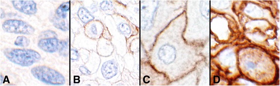
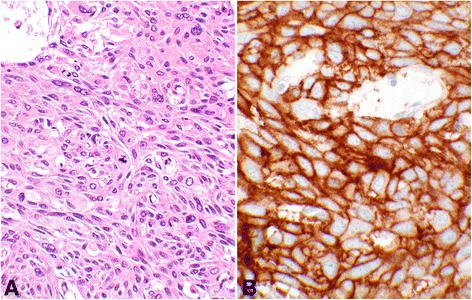
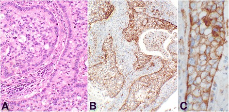
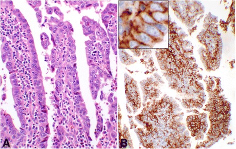
Methods: Immunohistochemical staining for PD-L1 expression was performed on a tissue microarray of 101 normal and neoplastic cervical tissues. Neoplastic cores were divided into three groups: squamous cell carcinoma, adenosquamous carcinoma, and endocervical adenocarcinoma. PD-L1 expression was scored based on an adopted scoring system accounting to percentage and intensity of positivity, and results provided alongside available clinical and demographic data.

Results: Overall, PD-L1 was positive in 32 of 93 (34.4%) cervical carcinomas. Subcategorically, PD-L1 was positive in 28 of 74 (37.8%) squamous cell carcinomas, two of seven (28.6%) adenosquamous carcinomas, and two of 12 (16.7%) endocervical adenocarcinomas. It was negative in six benign cervical tissues.

Conclusions: This study shows a significant expression of PD-L1 in 34.4% of cervical carcinomas and no expression of PD-L1 in benign cervical tissues. These findings suggest a role for further investigation of anti-PD-L1/PD-1 immunotherapies in the treatment of PD-L1-positive cervical tumors. In addition, our adopted scoring system will facilitate more systematic correlations between tumor reactivity and response to treatment.

Keywords: Adenosquamous carcinoma; Cervical cancer; Endocervical adenocarcinoma; Immunohistochemistry; Immunotherapy; PD-L1; Squamous cell carcinoma; Tissue microarray; Uterine cervix.


Figures

Fig. 1
Fig. 1
Intensities of the PD-L1 reactions (objective 60×). a Intensity 0, showing no cell membrane reaction (#8, Table 2). b Intensity 1+, showing weak and/or incomplete circumferential cell membrane reactions (#16, Table 2). c Intensity 2+, showing distinct complete circumferential cell membrane staining (#7, Table 1). d Intensity 3+, showing very strong circumferential cell membrane reaction (#47, Table 2)
Fig. 2
Fig. 2
Score 2 in squamous cell carcinoma. An example of Score 2b in grade-II squamous cell carcinoma of the cervix showing PD-L1 staining of 100% of the tumor cells (#49, Table 2). a shows hematoxylin and eosin stain of the carcinoma with moderate differentiation containing a mitotic figure at the center of the photomicrograph (objective 40×). b demonstrates the intense membrane reaction for PD-L1 with 3+ intensity involving all tumor cells (objective 40×)
Fig. 3
Fig. 3
Score 2 in adenosquamous carcinoma. An example of Score 2b adenosquamous carcinoma showing the PD-L1 reaction involving 100% of the tumor cells (#6, Table 4). a shows hematoxylin & eosin stain of the glandular organization of the tumor cells with adjacent stromal tissue. The center of the glandular structures contains solid nests of cells with squamoid differentiation (objective 20×). b demonstrates the PD-L1 membrane staining of all tumor cells (objective 10×). c shows complete circumferential cell membrane staining of the tumor cells with 2+ intensity (objective 40×). This intensity may be interpreted as 3+ by some other pathologists
Fig. 4
Fig. 4
Score 2 in endocervical adenocarcinoma. An example of Score 2b endocervical adenocarcinoma of the cervix with PD-L1 reaction involving 90% of the epithelial tumor cells (#12, Table 5). a shows the hematoxylin & eosin stain of the glandular structures with atypical epithelial cell lining and inflammatory stromal core (objective 20×). b demonstrates the membrane reaction for PD-L1 with 2+ intensity involving the epithelial tumor cells, many at cross section (objective 10×). The inset shows the circumferential membrane staining of the tumor cells for PD-L1 at a higher magnification (objective 60×)

References

    1. Reddy OL, Shintaku IP, Moatamed NA. Programmed cell death ligand-1 is expressed in a significant number of uterine cervical carcinomas. Mod Pathol. 2016;29(S2):305A. - PMC - PubMed
    1. Siegel RL, Miller KD, Jemal A. Cancer statistics, 2016. CA Cancer J Clin. 2016;66:7–30. doi: 10.3322/caac.21332. - DOI - PubMed
    1. Van Kriekinge G, Castellsague X, Cibula D, Demarteau N. Estimation of the potential overall impact of human papillomavirus vaccination on cervical cancer cases and deaths. Vaccine. 2014;32:733–739. doi: 10.1016/j.vaccine.2013.11.049. - DOI - PubMed
    1. NCCN. National Comprehensive Cancer Network guidlines in oncology: Cervical cancer. 2016. https://www.nccn.org/professionals/physician_gls/pdf/cervical.pdf. Accessed 21 Aug 2016.
    1. Sznol M, Chen L. Antagonist antibodies to PD-1 and B7-H1 (PD-L1) in the treatment of advanced human cancer. Clin Cancer Res. 2013;19:1021–1034. doi: 10.1158/1078-0432.CCR-12-2063. - DOI - PMC - PubMed