. 2017 Jun 16:7:200-210.
doi: 10.1016/j.omtn.2017.04.005. Epub 2017 Apr 12.

Evaluation of Antisense Oligonucleotides Targeting ATXN3 in SCA3 Mouse Models

Affiliations

Evaluation of Antisense Oligonucleotides Targeting ATXN3 in SCA3 Mouse Models

Lauren R Moore et al. Mol Ther Nucleic Acids. .

Abstract

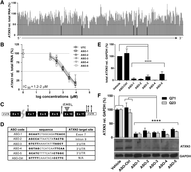
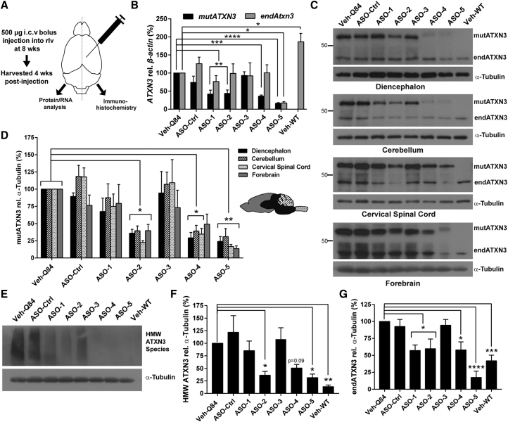
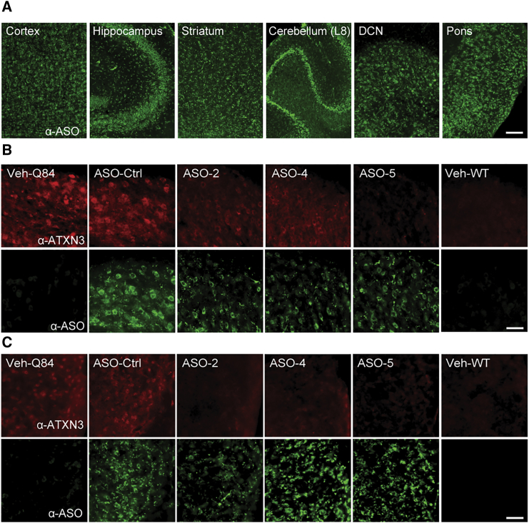
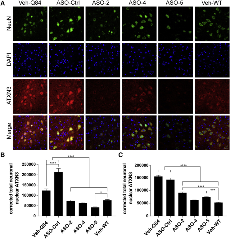
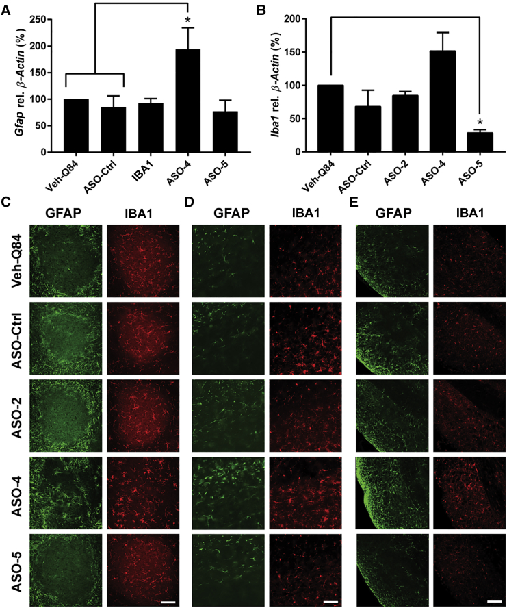
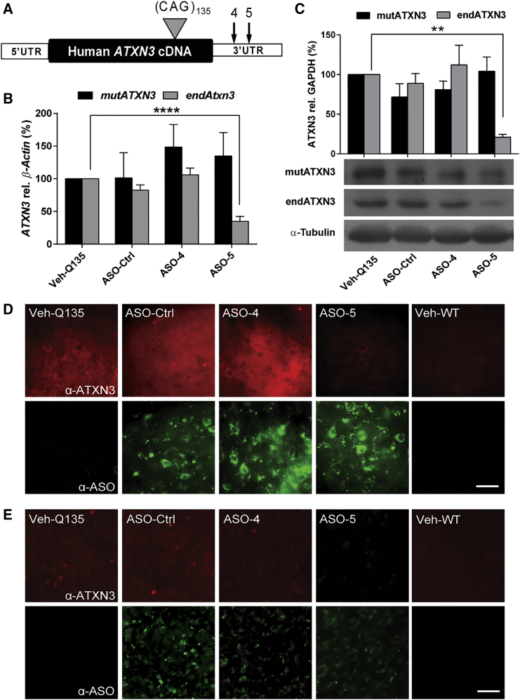
The most common dominantly inherited ataxia, spinocerebellar ataxia type 3 (SCA3), is an incurable neurodegenerative disorder caused by a CAG repeat expansion in the ATXN3 gene that encodes an abnormally long polyglutamine tract in the disease protein, ATXN3. Mice lacking ATXN3 are phenotypically normal; hence, disease gene suppression offers a compelling approach to slow the neurodegenerative cascade in SCA3. Here we tested antisense oligonucleotides (ASOs) that target human ATXN3 in two complementary mouse models of SCA3: yeast artificial chromosome (YAC) MJD-Q84.2 (Q84) mice expressing the full-length human ATXN3 gene and cytomegalovirus (CMV) MJD-Q135 (Q135) mice expressing a human ATXN3 cDNA. Intracerebroventricular injection of ASOs resulted in widespread delivery to the most vulnerable brain regions in SCA3. In treated Q84 mice, three of five tested ASOs reduced disease protein levels by >50% in the diencephalon, cerebellum, and cervical spinal cord. Two ASOs also significantly reduced mutant ATXN3 in the mouse forebrain and resulted in no signs of astrogliosis or microgliosis. In Q135 mice expressing a single ATXN3 isoform via a cDNA transgene, ASOs did not result in similar robust ATXN3 silencing. Our results indicate that ASOs targeting full-length human ATXN3 would likely be well tolerated and could lead to a preventative therapy for SCA3.

Keywords: ASO; ATXN3; MJD; Machado-Joseph disease; SCA3; antisense oligonucleotide; polyglutamine disease; spinocerebellar ataxia type 3.


Figures

Figure 1
Figure 1
Identification of Human ATXN3 Antisense Oligonucleotides (A) Screen of ASOs complementary to human ATXN3 in HEPG2 cells. 2 μM ASO was electroporated into HEPG2 cells; 24 hr post-treatment, ATXN3 mRNA levels were quantified by qPCR and normalized to total RNA levels. Data expressed as the percent of untransfected control cells. ASOs listed in order of relative binding site on ATXN3 transcript (5′ to 3′). Five ASOs characterized further are noted in black. (B) Dose response of top five ASOs in HEPG2 cells. IC50s calculated from four-point non-linear fit dose-response curve. (C) Schematic of anti-ATXN3 ASO target sites on the human ATXN3 transcript. (D) Anti-ATXN3 ASOs and the non-specific ASO-Ctrl nucleotide sequence possess a 5-8-5 MOE gapmer design in which an 8-mer block of unmodified deoxynucleotides is flanked by 5-mer blocks of 2′-O-methoxyethyl (MOE)-modified ribonucleotides indicated in bold. (E) Total ATXN3 transcript levels in SCA3 patient fibroblasts 48 hr after transfection with anti-ATXN3 ASOs (4 μM), ASO-Ctrl, or vehicle. Data (mean ± SEM) are reported relative to fibroblasts treated with vehicle alone (n = 6 per group). One-way ANOVA statistical analysis performed with the post hoc Tukey test (****p < 0.0001). (F) Immunoblotting and quantification of expanded (Q71) and wild-type (Q23) ATXN3 protein in SCA3 patient fibroblasts 72 hr after transfection with anti-ATXN3 ASOs, ASO-Ctrl, or vehicle. Data (mean ± SEM) are reported relative to fibroblasts treated with vehicle (n = 6 per group). One-way ANOVA performed with the post hoc Tukey test (****p < 0.0001).
Figure 2
Figure 2
In Vivo Suppression of Mutant ATXN3 by Anti-ATXN3 ASOs in Q84 Mice, a YAC Transgenic Mouse Model of SCA3 (A) Schematic of anti-ATXN3 ASO trial design. Sex-matched hemizygous Q84 mice received a single i.c.v. bolus injection of 500 μg ASO or vehicle into the right lateral ventricle (rlv) at 8 weeks of age. Brains were harvested and dissected 4 weeks later for RNA, protein, and immunohistochemical analysis. (B) Quantification of endogenous Atxn3 (endAtxn3) and mutant ATXN3 (mutATXN3) transcripts in the diencephalon of vehicle- (Veh-Q84 and Veh-WT), ASO-Ctrl-, and anti-ATXN3 ASO-treated mice. (C) Representative western blots of mutant ATXN3 (mutATXN3) and endogenous ATXN3 (endATXN3) protein expression in major brain regions of treated mice. (D) Quantification of mutATXN3 protein expression in major brain regions of treated mice. (E and F) Representative western blot (E) and quantification of high molecular weight (HMW) ATXN3 species in the diencephalon of treated mice (F). (G) Quantification of endATXN3 protein expression in the diencephalon of treated mice. Data (mean ± SEM) are reported relative to mice treated with vehicle (n = 6 per group). One-way ANOVA performed with the post hoc Dunnett test (*p < 0.05; **p < 0.01; ***p < 0.001; ****p < 0.0001). WT, wild type.
Figure 3
Figure 3
Anti-ATXN3 ASOs Distribute Widely throughout the CNS and Suppress ATXN3 Protein Expression in Q84 Mice (A) Representative immunofluorescence images of ASO (green) distributed throughout key SCA3-affected brain regions in Q84 hemizygous mice 4 weeks after injection. (B and C) Representative immunofluorescence images of ASO-mediated (green) suppression of ATXN3 (red) in deep cerebellar nuclei (DCN) (B) and pontine nuclei (C) of Q84 hemizygous mice 4 weeks post-treatment. Scale bars represent 200 μm in (A) and 50 μm in (B) and (C).
Figure 4
Figure 4
ASOs Significantly Suppress ATXN3 Accumulation within Neuronal Nuclei (A) Representative immunofluorescence images of ATXN3 (red) reduction within neuronal nuclei (NeuN, green; DAPI, blue) in the deep cerebellar nuclei (DCN) of Q84 hemizygous mice, 4 weeks post-treatment. Scale bar represents 25 μm. (B and C) Quantification of total corrected neuronal nuclear ATXN3 fluorescence in the DCN (B) and pontine nuclei (C). Data (mean ± SEM) are reported relative to Q84 vehicle-treated mice (n = 3 per group). One-way ANOVA performed with the post hoc Tukey test (*p < 0.05; ***p < 0.001; ****p < 0.0001).
Figure 5
Figure 5
ASOs Result in Limited Immunoreactive Changes in Q84 Mice (A and B) Transcript levels of the astrocytic marker Gfap (A) and microglial marker Iba1 (B) in the left diencephalon of vehicle and ASO-treated mice 4 weeks after injection. Means ± SEM are reported relative to Q84 vehicle-treated mice (n = 6 per group). One-way ANOVA statistical analysis performed with the post hoc Tukey test (*p < 0.05). (C–E) Representative GFAP (green) and IBA1 (red) immunofluorescence images of the deep cerebellar nuclei (DCN) (C), cerebral cortex (D), and body of the pons (E). Scale bars represent 100 μm in (C) and (D) and 50 μm in (E).
Figure 6
Figure 6
ASOs Do Not Reduce Mutant ATXN3 Expression in a Second Model, the Q135 cDNA Transgenic Mouse, Despite Effective Delivery (A) Schematic of anti-ATXN3 ASO target sites on the CMV MJD-Q135 ATXN3 cDNA transcript. (B) Mutant ATXN3 (mutATXN3) and endogenous Atxn3 (endAtxn3) transcript levels in the diencephalon of Q135 mice 4 weeks after injection (n = 6 per group). Data shown are mean ± SEM relative to Q135 vehicle-treated mice (n = 6 per group). One-way ANOVA statistical analysis performed with the post hoc Dunnett test (∗∗p < 0.01; ∗∗∗∗p < 0.0001). (C) Western blotting and quantification of mutant human ATXN3 and endogenous murine ATXN3 expression in Q135 diencephalon 4 weeks after injection. (D and E) Representative ATXN3 (red) and ASO (green) immunofluorescence images of the deep cerebellar nuclei (DCN) (D) and pontine nuclei (E). Scale bars represent 50 μm.

References

    1. Costa Mdo.C., Paulson H.L. Toward understanding Machado-Joseph disease. Prog. Neurobiol. 2012;97:239–257. - PMC - PubMed
    1. Alves S., Nascimento-Ferreira I., Auregan G., Hassig R., Dufour N., Brouillet E., Pedroso de Lima M.C., Hantraye P., Pereira de Almeida L., Déglon N. Allele-specific RNA silencing of mutant ataxin-3 mediates neuroprotection in a rat model of Machado-Joseph disease. PLoS ONE. 2008;3:e3341. - PMC - PubMed
    1. Costa Mdo.C., Luna-Cancalon K., Fischer S., Ashraf N.S., Ouyang M., Dharia R.M., Martin-Fishman L., Yang Y., Shakkottai V.G., Davidson B.L. Toward RNAi therapy for the polyglutamine disease Machado-Joseph disease. Mol. Ther. 2013;21:1898–1908. - PMC - PubMed
    1. Rodriguez-Lebron E., Costa Mdo C., Luna-Cancalon K., Peron T.M., Fischer S., Boudreau R.L., Davidson B.L., Paulson H.L. Silencing mutant ATXN3 expression resolves molecular phenotypes in SCA3 transgenic mice. Mol. Ther. 2013;21:1909–1918. - PMC - PubMed
    1. Nóbrega C., Nascimento-Ferreira I., Onofre I., Albuquerque D., Hirai H., Déglon N., de Almeida L.P. Silencing mutant ataxin-3 rescues motor deficits and neuropathology in Machado-Joseph disease transgenic mice. PLoS ONE. 2013;8:e52396. - PMC - PubMed