. 2017 Sep:140:45-57.
doi: 10.1016/j.biomaterials.2017.06.010. Epub 2017 Jun 9.

Substrate stiffness and VE-cadherin mechano-transduction coordinate to regulate endothelial monolayer integrity

Affiliations

Substrate stiffness and VE-cadherin mechano-transduction coordinate to regulate endothelial monolayer integrity

Roberto C Andresen Eguiluz et al. Biomaterials. 2017 Sep.

Abstract

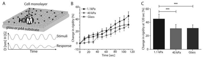
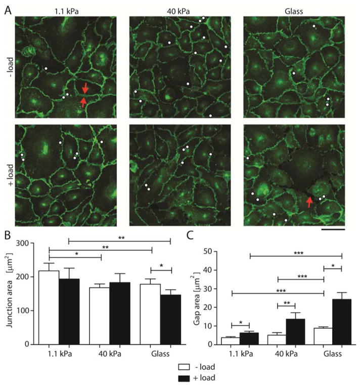
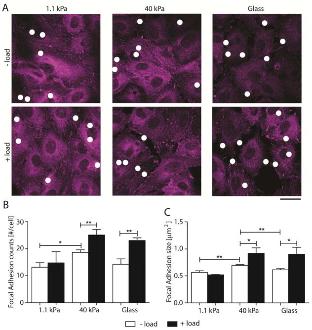
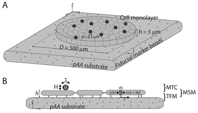
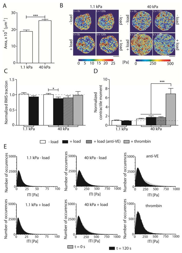
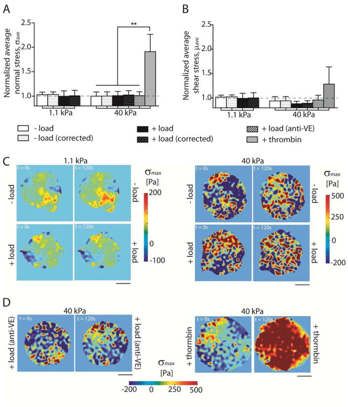
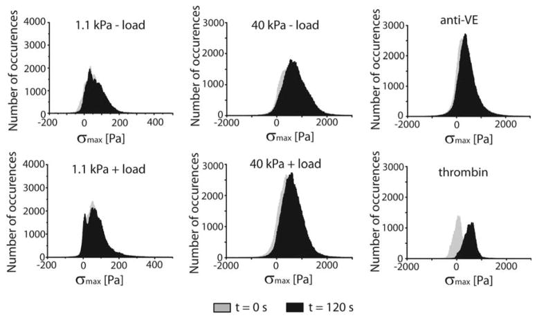
The vascular endothelium is subject to diverse mechanical cues that regulate vascular endothelial barrier function. In addition to rigidity sensing through integrin adhesions, mechanical perturbations such as changes in fluid shear stress can also activate force transduction signals at intercellular junctions. This study investigated how extracellular matrix rigidity and intercellular force transduction, activated by vascular endothelial cadherin, coordinate to regulate the integrity of endothelial monolayers. Studies used complementary mechanical measurements of endothelial monolayers grown on patterned substrates of variable stiffness. Specifically perturbing VE-cadherin receptors activated intercellular force transduction signals that increased integrin-dependent cell contractility and disrupted cell-cell and cell-matrix adhesions. Further investigations of the impact of substrate rigidity on force transduction signaling demonstrated how cells integrate extracellular mechanics cues and intercellular force transduction signals, to regulate endothelial integrity and global tissue mechanics. VE-cadherin specific signaling increased focal adhesion remodeling and cell contractility, while sustaining the overall mechanical equilibrium at the mesoscale. Conversely, increased substrate rigidity exacerbates the disruptive effects of intercellular force transduction signals, by increasing heterogeneity in monolayer stress distributions. The results provide new insights into how substrate stiffness and intercellular force transduction coordinate to regulate endothelial monolayer integrity.

Keywords: Cell traction; Magnetic twisting cytometry; Mechanotransduction; Micropatterned substrates; VE-cadherin.


Figures

Fig. 1
Fig. 1. VE-cadherin mediated adaptive cell stiffening depends on substrate rigidity
(A) Schematic of the MTC showing magnetized beads (M), oscillating magnetic field (H), and resulting twisting torque (τ) that displaces beads. Bead displacement amplitudes reveal the viscoelastic behavior of the bead-cell junction. All beads were functionalized with VE-cadherin-Fc. (B) Time dependence of force actuated cell stiffening relative to the initial basal value. Endothelial monolayers grown on soft (1.1 kPa) and stiff (40 kPa) pAA hydrogels, and glass (50 GPa, blue) substrates were perturbed with VE-cadherin coated beads for 120 sec. (C) Change in stiffness after perturbation, relative to basal values. Data represents mean ± s.d. (n = 212 for glass, n = 202 for 40 kPa, and n = 181 for 1.1 kPa beads, *** p < 0.001).
Fig. 2
Fig. 2. Substrate stiffness modulates intercellular junction remodeling following VE-cadherin-mediated mechanotransduction
(A) Confocal immunofluorescence images of EC monolayers bound with VE-cad-Fc beads without (− load) and with (+ load) applied perturbation. Scale bar = 50 μm. The immunofluorescence images show VE-cadherin distributions on 1.1 kPa, 40 kPa, and glass (~50 GPa) substrata. White dots indicate locations of VE-cadherin coated beads. Red arrows show junction area and gap. Images represent > 10 images per condition from 3 independent experiments. (B) and (C) Quantification of interendothelial junction and gap areas between endothelial cells without and with oscillating shear stress for 120 sec (n = 100–150 cells from 3 independent experiments). Data show the mean ± s.e.m. * p < 0.05, ** p < 0.01, *** p < 0.001.
Fig. 3
Fig. 3. VE-cadherin mechanotransduction alters number and size of focal adhesion
(A) Confocal images of immunofluorescence EC monolayers showing focal adhesions without (− load) and with (+ load) VE-cadherin specific perturbations. FA were stained for paxillin. Scale bar = 25 μm. White dots indicate location of VE-cadherin coated beads. (B) and (C) Quantification of FA number per cell and size, respectively, without (white bars) and with (black bars) oscillating shear stress for 120 sec (n = 70 – 100 cells over 2 independent experiments). Data show the mean ± s.e.m. * p < 0.05, ** p < 0.01.
Fig. 4
Fig. 4. Integrated platform consisting of MTC + TFM + MSM to quantify stresses due to VE-cadherin mechanotransduction in EC monolayers
(A) FN arrays 500 μm in diameter on pAA substrates with embedded fiducial marker beads were seeded with HPAECs for 48 hrs, yielding regular circular cell colonies with 200–280 cells and 5 μm height. (B) Schematics showing the applied stresses (MTC), recovered stresses (tractions), and calculated stresses (MSM).
Fig. 5
Fig. 5. VE-cadherin mechanotransduction decreases effective cell-substrate area interactions on stiff substrates
(A) Projected area of EC colonies plated on 1.1 kPa and 40 kPa. (B) Representative heat maps showing tractions at time points t = 0 sec and t = 120 sec for soft and stiff hydrogels without (− load) and with force loading (+ load). Scale bars = 150 μm, showing the heterogeneous spatial tractions distribution. (C) Normalized RMS traction of cell colonies. (D) Normalized NCM. (E) Frequency histograms of the tractions magnitude showing the tractions magnitude distribution, ITI at time points t = sec (grey) and t = 120 sec (black). Data show the mean ± s.e.m. n = 10, 11, 10, 11, 11, and 3 for the 1.1 kPa − load, 1.1 kPa + load, 40 kPa − load, 40 kPa + load, 40 kPa + load (anti-VE), and + thrombin, respectively. * p < 0.05, *** p < 0.0001.
Fig. 6
Fig. 6. Mechanical integrity of EC colonies is maintained even with VE-cadherin mechanotransduction
(A) Normalized σave, ± load and (B) normalized μave, ± load. (C) Representative heat maps showing monolayer σmax distributions at time points t = 0 sec and t = 120 sec for soft and stiff hydrogels. (D) Representative heat maps showing σmax for same conditions as (A) for negative (anti-VE) and positive (thrombin) controls. Data show the mean ± s.e.m. n = 10, 11, 10, 11, 11, and 3 for the 1.1 kPa − load, 1.1 kPa + load, 40 kPa − load, 40 kPa + load, 40 kPa + load (anti-VE), and + thrombin, respectively. ** p < 0.005.
Fig. 7
Fig. 7. Frequency histograms
σmax vectors at timepoints t = 0 sec (grey) and t = 120 sec (black). n = 10, 11, 10, 11, 11, and 3 for the 1.1 kPa − load, 1.1 kPa + load, 40 kPa − load, 40 kPa + load, 40 kPa + load (anti-VE), and + thrombin, respectively.

References

    1. Tzima E, Irani-Tehrani M, Kiosses WB, DeJana E, Schultz Da, Engelhardt B, Cao G, DeLisser H, Schwartz Ma. A mechanosensory complex that mediates the endothelial cell response to fluid shear stress. Nature. 2005;437:426–431. doi: 10.1038/Nature03952. - DOI - PubMed
    1. Kohn JC, Zhou DW, Bordeleau F, Zhou AL, Mason BN, Mitchell MJ, King MR, Reinhart-King CA. Cooperative effects of matrix stiffness and fluid shear stress on endothelial cell behavior. Biophys J. 2015;108:471–478. doi: 10.1016/j.bpj.2014.12.023. - DOI - PMC - PubMed
    1. Birukova AA, Chatchavalvanich S, Rios A, Kawkitinarong K, Garcia JGN, Birukov KG. Differential regulation of pulmonary endothelial monolayer integrity by varying degrees of cyclic stretch. Am J Pathol. 2006;168:1749–1761. doi: 10.2353/ajpath.2006.050431. - DOI - PMC - PubMed
    1. Dan A, Huang RB, Leckband DE. Dynamic Imaging Reveals Coordinate Effects of Cyclic Stretch and Substrate Stiffness on Endothelial Integrity. Ann Biomed Eng. 2016:1–13. doi: 10.1007/s10439-016-1677-4. - DOI - PMC - PubMed
    1. Cummins PM, Sweeney NVO, Killeen MT, Birney YA, Redmond EM, Cahill PA. Cyclic strain-mediated matrix metalloproteinase regulation within the vascular endothelium: a force to be reckoned with. Am J Physiol Hear Circ Physiol. 2007;292:H28–H42. doi: 10.1152/ajpheart.00304.2006. - DOI - PubMed