Review
. 2017 Nov;74(22):4077-4095.
doi: 10.1007/s00018-017-2570-5. Epub 2017 Jun 17.

Routes and machinery of primary cilium biogenesis

Affiliations
Review

Routes and machinery of primary cilium biogenesis

Miguel Bernabé-Rubio et al. Cell Mol Life Sci. 2017 Nov.

Abstract

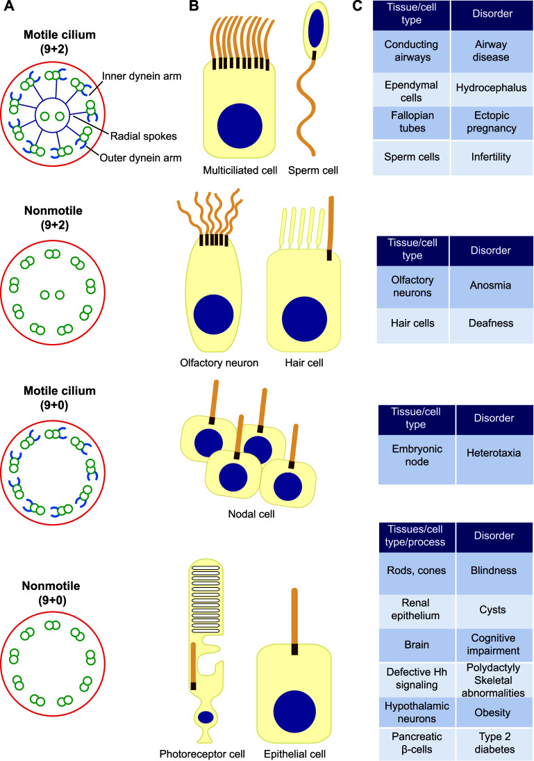
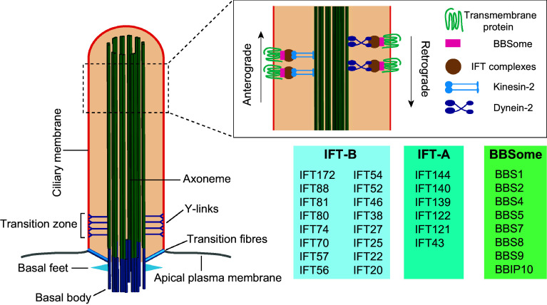
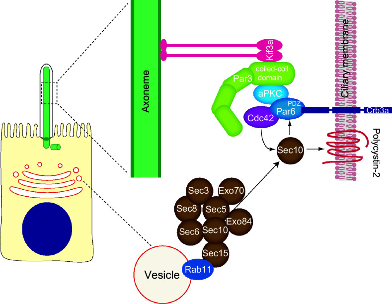
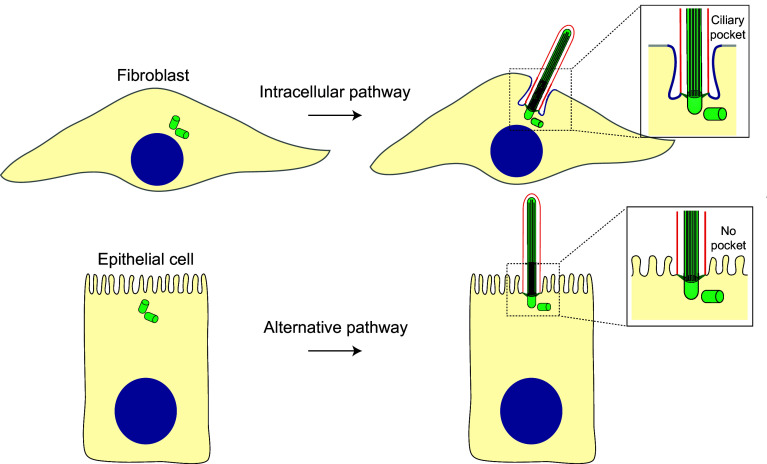
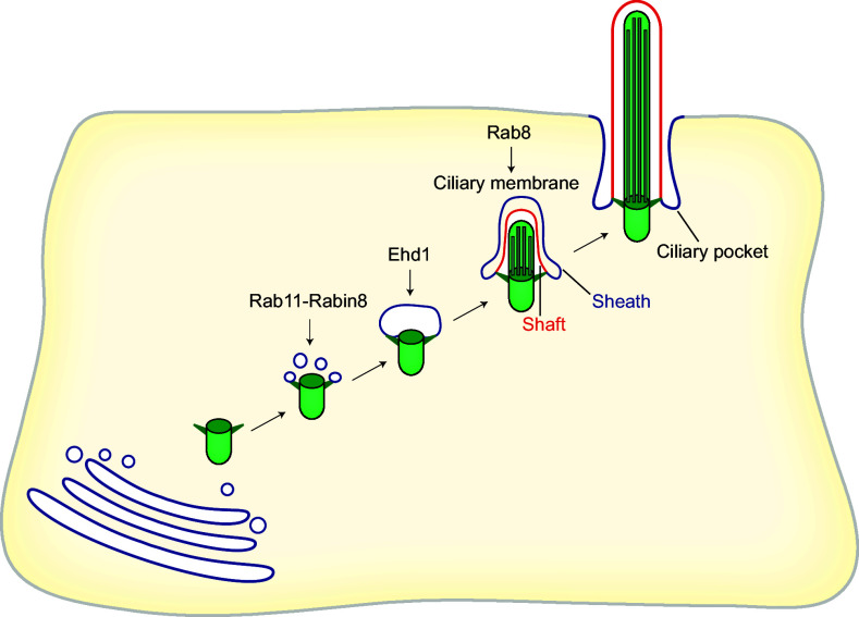
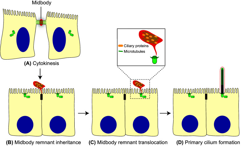
Primary cilia are solitary, microtubule-based protrusions of the cell surface that play fundamental roles as photosensors, mechanosensors and biochemical sensors. Primary cilia dysfunction results in a long list of developmental and degenerative disorders that combine to give rise to a large spectrum of human diseases affecting almost any major body organ. Depending on the cell type, primary ciliogenesis is initiated intracellularly, as in fibroblasts, or at the cell surface, as in renal polarized epithelial cells. In this review, we have focused on the routes of primary ciliogenesis placing particular emphasis on the recently described pathway in renal polarized epithelial cells by which the midbody remnant resulting from a previous cell division event enables the centrosome for initiation of primary cilium assembly. The protein machinery implicated in primary cilium formation in epithelial cells, including the machinery best known for its involvement in establishing cell polarity and polarized membrane trafficking, is also discussed.

Keywords: Alternative pathway; Fibroblasts; Intracellular pathway; Midbody remnant; Polarized epithelial cells; Primary ciliogenesis; Protein machinery.


Figures

Fig. 1
Fig. 1
Types of cilium. a Schematic of distinct types of cilium as seen in cross-section. b Examples of cell types harboring each type of cilium. c Examples of disorders associated with the dysfunction of the distinct types of cilium
Fig. 2
Fig. 2
General structure of the primary cilium. The basal body is attached to the ciliary membrane by the transition fibers. The axoneme constitutes the backbone of the cilium and is surrounded by the ciliary membrane, which is continuous with the plasma membrane. IFT is carried out by the IFT-A and IFT-B complexes powered by dynein-2 and kinesin-2 motors, respectively, and with the participation of the BBSome complex
Fig. 3
Fig. 3
Multi-protein complexes involved in polarized trafficking and cell polarity implicated in primary cilium formation by polarized epithelial cells. In addition to their role in polarization process, the exocyst and the Par complexes participate in primary cilium assembly. Par complex consists of Par3, Par6, aPKC and Cdc42. The motor Kif3a targets the Par complex to the ciliary axoneme through interaction with the coiled-coil domain of Par3. The Par complex has been proposed to be an adaptor for targeting the transmembrane protein Crumbs3a to the ciliary membrane through association with the Par6 PDZ domain. Cdc42 is responsible for recruitment of exocyst complex, which targets important cargo, such as polycystin-2, to the cilium
Fig. 4
Fig. 4
Routes of primary ciliogenesis. The position of the centrosome, near the nucleus or close to the plasma membrane, and the presence or absence of a ciliary pocket predicts the type of pathway used for primary ciliogenesis. Fibroblasts and polarized epithelial cells are shown as examples of cells that use the intracellular and alternative routes, respectively
Fig. 5
Fig. 5
The intracellular pathway. Ciliogenesis initiates intracellularly with the formation of a large ciliary vesicle at the distal end of the appendages of the mother centriole by fusion of smaller vesicles. The axoneme starts forming intracellularly and, as it grows, deforms the ciliary vesicle and establishes an inner membrane (shaft) and an outer membrane (sheath). The incipient cilium is finally exocytosed and the cilium becomes exposed in the plasma membrane. The sheath gives rise to the ciliary pocket, and the shaft forms the ciliary membrane
Fig. 6
Fig. 6
The alternative route. a In polarized epithelial cells, the intercellular bridge containing ciliary proteins forms at the apical cell surface during cytokinesis. b When abscission occurs, one of the two daughter cells inherits the midbody remnant, which localizes apically at the cell periphery, near the tight junctions. c The remnant subsequently moves over the apical surface towards the centrosome, which is docked at the center of the apical membrane. d When the midbody meets the centrosome, the initiation of primary cilium assembly is facilitated. The entire process of primary cilium formation takes place in the plasma membrane

References

    1. Goetz SC, Anderson KV. The primary cilium: a signalling centre during vertebrate development. Nat Rev Genet. 2010;11:331–344. doi: 10.1038/nrg2774. - DOI - PMC - PubMed
    1. Satir P, Christensen ST. Overview of structure and function of mammalian cilia. Annu Rev Physiol. 2007;69:377–400. doi: 10.1146/annurev.physiol.69.040705.141236. - DOI - PubMed
    1. Ishikawa H, Marshall WF. Ciliogenesis: building the cell’s antenna. Nat Rev Mol Cell Biol. 2011;12:222–234. doi: 10.1038/nrm3085. - DOI - PubMed
    1. Brooks ER, Wallingford JB. Multiciliated cells: a review. Curr Biol. 2014;24:R973–R982. doi: 10.1016/j.cub.2014.08.047. - DOI - PMC - PubMed
    1. Lindemann CB, Lesich KA. Functional anatomy of the mammalian sperm flagellum. Cytoskeleton. 2016;73:652–669. doi: 10.1002/cm.21338. - DOI - PubMed

Publication types

Substances

LinkOut - more resources